Hostname: page-component-586b7cd67f-t8hqh Total loading time: 0 Render date: 2024-11-27T01:41:11.913Z Has data issue: false hasContentIssue false

Structure of the Ebola Virus Nucleocapsid Core by Single Particle Cryo-Electron Microscopy

Published online by Cambridge University Press:  25 July 2016

Yukihiko Sugita
Affiliation:
Okinawa Institute of Science and Technology Graduate University, Onna, Japan
Yoshihiro Kawaoka
Affiliation:
Institute of Medical Science,University of Tokyo, Tokyo, Japan School of Veterinary Medicine, University of Wisconsin-Madison, Wisconsin, USA
Takeshi Noda
Affiliation:
Institute for Virus Research, Kyoto University, Kyoto, Japan
Matthias Wolf
Affiliation:
Okinawa Institute of Science and Technology Graduate University, Onna, Japan

Abstract

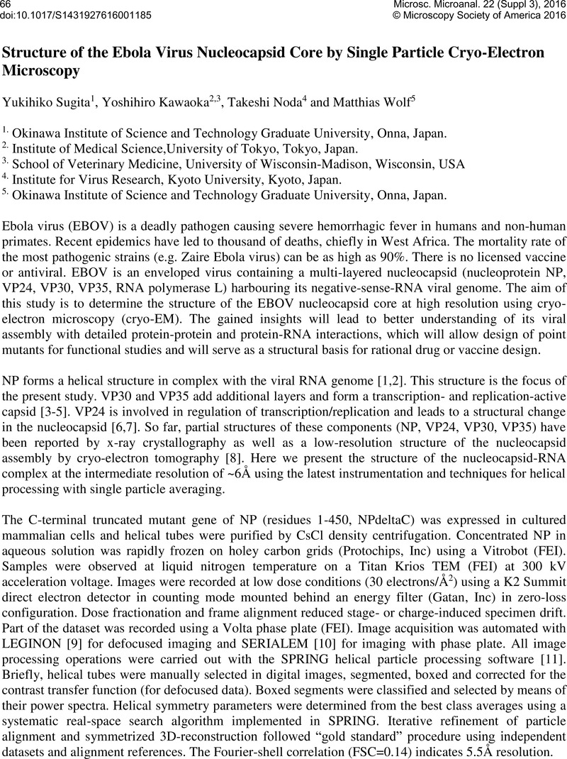
Image of the first page of this content. For PDF version, please use the ‘Save PDF’ preceeding this image.'
Type
Abstract
Copyright
© Microscopy Society of America 2016 

References

[1] Noda, , et al, PLoS Pathog 2(9) e99 2006 864.Google Scholar
[2] Noda, , et al, J Gen Virol 91(Pt6 (2010) 1478.Google Scholar
[3] Mühlberger, , et al, J Virol 73(3 (1999) 23332342.Google Scholar
[4] Halfmann, , et al, PNAS 105 (2008) 1129.Google Scholar
[5] Noda, , et al, J Infect Dis 204(Supp3 (2011) S878.Google Scholar
[6] Noda, , et al, J Infect Dis 196(Supp2 (2007) S247.Google Scholar
[7] Watanabe, , et al, J Infect Dis 196(Supp2 (2007) S284.Google Scholar
[8] Bharat, , et al, PNAS ( ) 109 11 (2012) 4275.Google Scholar
[9] Suloway, , et al, J Struct Biol 151(1 (2005) 41.Google Scholar
[10] Mastronarde, DN J Struct Biol 152(1 (2005) 36.Google Scholar
[11] Desfosses, , et al, J Struct Biol 185(1 (2014) 15.CrossRefGoogle Scholar
[12] The authors are grateful to R. Danev for advice on phase plate imaging and help with the setup of SerialEM. We thank OIST and the Japanese Government for generous research support.Google Scholar