Article contents
Soft X-Ray and Cathodoluminescence Examination of a Tanzanian Graphite Deposit
Published online by Cambridge University Press: 06 April 2020
Abstract

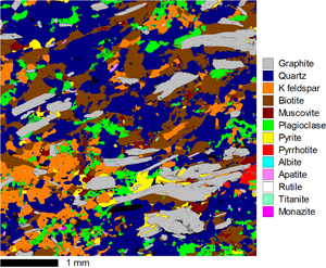
Hyperspectral soft X-ray emission (SXE) and cathodoluminescence (CL) spectrometry have been used to investigate a carbonaceous-rich geological deposit to understand the crystallinity and morphology of the carbon and the associated quartz. Panchromatic CL maps show both the growth of the quartz and the evidence of recrystallization. A fitted CL map reveals the distribution of Ti4+ within the grains and shows subtle growth zoning, together with radiation halos from 238U decay. The sensitivity of the SXE spectrometer to carbon, together with the anisotropic X-ray emission from highly orientated pyrolytic graphite, has enabled the C Kα peak shape to be used to measure the crystal orientation of individual graphite regions. Mapping has revealed that most grains are predominantly of a single orientation, and a number of graphite grains have been investigated to demonstrate the application of this new SXE technique. A peak fitting approach to analyzing the SXE spectra was developed to project the C Kα 2pz and 2p(x+y) orbital components of the graphite. The shape of these two end-member components is comparable to those produced by electron density of states calculations. The angular sensitivity of the SXE spectrometer has been shown to be comparable to that of electron backscatter diffraction.
- Type
- Australian Microbeam Analysis Society Special Section AMAS XV 2019
- Information
- Copyright
- Copyright © Microscopy Society of America 2020
References
- 2
- Cited by


