Hostname: page-component-78c5997874-8bhkd Total loading time: 0 Render date: 2024-11-03T01:26:52.023Z Has data issue: false hasContentIssue false

Quantitative STEM-EELS Imaging of Ferritin Distributions in Cultured Erythroblasts Undergoing Erythropoiesis

Published online by Cambridge University Press:  01 August 2018

M.A. Aronova
Affiliation:
National Institute of Biomedical Imaging and Bioengineering, NIH, Bethesda, MD
C. Byrnes
Affiliation:
National Institute of Diabetes and Digestive and Kidney Diseases, NIH, BethesdaMD
Y.T. Lee
Affiliation:
National Institute of Diabetes and Digestive and Kidney Diseases, NIH, BethesdaMD
G. Zhang
Affiliation:
National Institute of Biomedical Imaging and Bioengineering, NIH, Bethesda, MD
Y.C. Kim
Affiliation:
Center for Materials Physics and Technology, NRL, Washington, DC
E.R. Meier
Affiliation:
National Institute of Diabetes and Digestive and Kidney Diseases, NIH, BethesdaMD Children's National Health System, Washington, DC
R.D. Leapman
Affiliation:
National Institute of Biomedical Imaging and Bioengineering, NIH, Bethesda, MD

Abstract

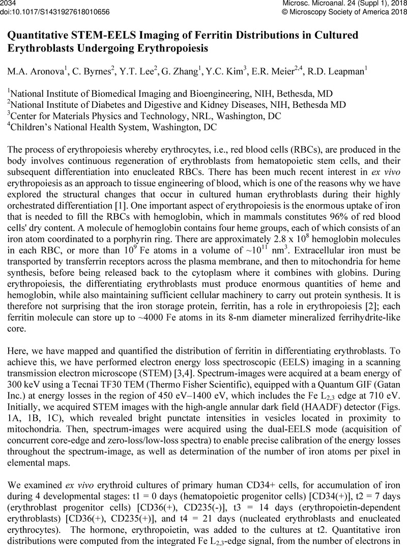
Image of the first page of this content. For PDF version, please use the ‘Save PDF’ preceeding this image.'
Type
Abstract
Copyright
© Microscopy Society of America 2018 

References

[1] van Veen, T Hunt, JA Tissue Engin. Regen., Med. 9 2015) p. 760.Google Scholar
[2] Vaisman, B, Fibach, E Konijn, AM Blood 90 1997) p. 831.Google Scholar
[3] Leapman, RD J. Microsc. 210 2003) p. 5.Google Scholar
[4] Fukunaga, M, et al, Proc. Natl. Acad. Sci. USA 107 2010) p. 3834.Google Scholar
[5] This research was supported by the intramural programs of NIBIB and NIDDK at the NIH.Google Scholar