Hostname: page-component-586b7cd67f-gb8f7 Total loading time: 0 Render date: 2024-11-26T16:20:22.174Z Has data issue: false hasContentIssue false

Quantitative Correlative Light and Electron Microscopies; Targeting the Host Actin Cytoskeleton

Published online by Cambridge University Press:  27 August 2014

Niels Volkmann
Affiliation:
Bioinformatics and Structural Biology Program, Sanford Burnham Medical Research Institute, La Jolla, USA
Dorit Hanein
Affiliation:
Bioinformatics and Structural Biology Program, Sanford Burnham Medical Research Institute, La Jolla, USA

Abstract

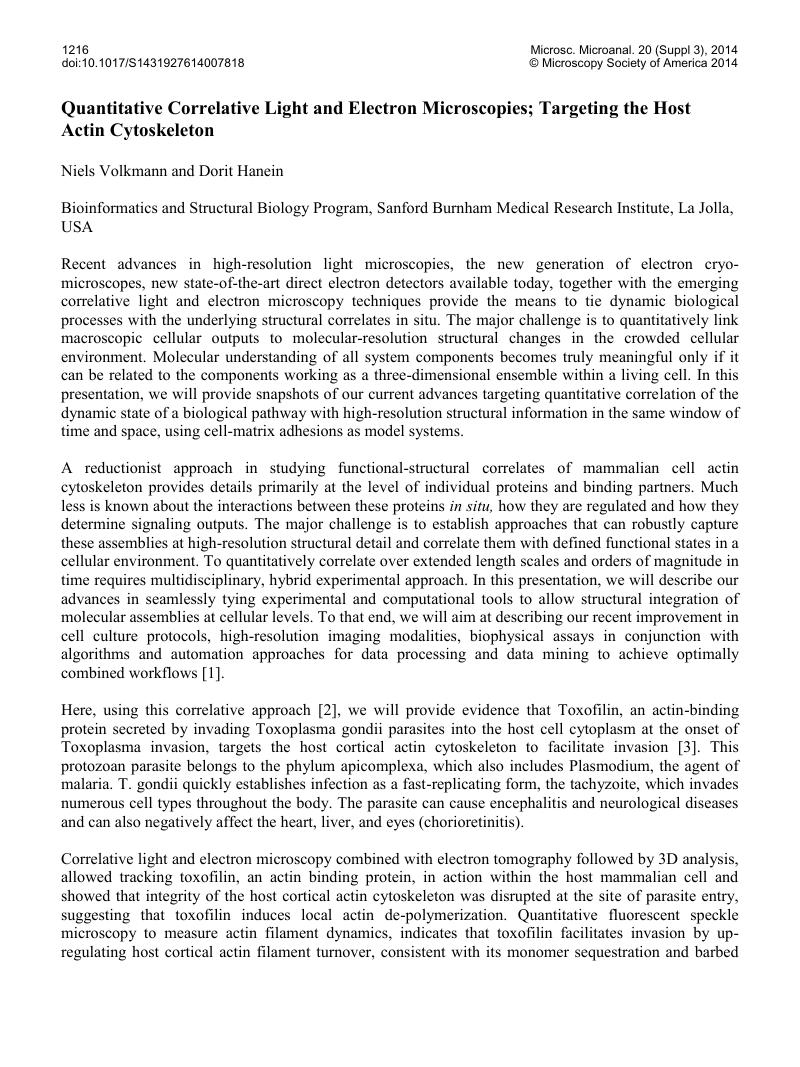
Image of the first page of this content. For PDF version, please use the ‘Save PDF’ preceeding this image.'
Type
Abstract
Copyright
Copyright © Microscopy Society of America 2014 

References

[1] These studies are supported by the National Institute of General Sciences (NIGMS) Grant Number P01 GM098412 (DH and NV).Google Scholar
[2] Hanein, D, Volkmann, N. Correlative light-electron microscopy. Adv Protein Chem Struct Biol. 82, 91-99 (2011).Google Scholar
[3] Delorme-Walker, V, Abrivard, M, Lagal, V, Anderson, K, Perazzi, A, Gonzalez, V, Page, C, Chauvet, J, Ochoa, W, Volkmann, N, Hanein, D, and Tardieux, I. Toxofilin upregulates the host cortical actin cytoskeleton dynamics, facilitating Toxoplasma invasion. J Cell Sci. 125, 4333-4342 (2012).Google Scholar
[4] Lee, S. H., Hayes, D. B., Rebowski, G., Tardieux, I. and Dominguez, R. Toxofilin from Toxoplasma gondii forms a ternary complex with an antiparallel actin dimer. Proc. Natl. Acad. Sci. USA 104, 16122–16127 (2007).Google Scholar