Hostname: page-component-cd9895bd7-gbm5v Total loading time: 0 Render date: 2025-01-03T16:56:36.518Z Has data issue: false hasContentIssue false

Precisely Picking Nanoparticles by a “Nano-Scalpel” for 360° Electron Tomography

Published online by Cambridge University Press:  14 September 2022
Xiaohui Huang
Affiliation:
Institute of Nanotechnology, Karlsruhe Institute of Technology, Hermann-von-Helmholtz-Platz 1, 76344 Eggenstein-Leopoldshafen, Germany Department of Materials and Earth Sciences, Technical University of Darmstadt, Alarich-Weiss-Straße 2, 64287 Darmstadt, Germany
Yushu Tang*
Affiliation:
Institute of Nanotechnology, Karlsruhe Institute of Technology, Hermann-von-Helmholtz-Platz 1, 76344 Eggenstein-Leopoldshafen, Germany
Christian Kübel
Affiliation:
Institute of Nanotechnology, Karlsruhe Institute of Technology, Hermann-von-Helmholtz-Platz 1, 76344 Eggenstein-Leopoldshafen, Germany Department of Materials and Earth Sciences, Technical University of Darmstadt, Alarich-Weiss-Straße 2, 64287 Darmstadt, Germany Karlsruhe Nano Micro Facility (KNMF), Karlsruhe Institute of Technology, Hermann-von-Helmholtz-Platz 1, 76344 Eggenstein-Leopoldshafen, Germany
Di Wang*
Affiliation:
Institute of Nanotechnology, Karlsruhe Institute of Technology, Hermann-von-Helmholtz-Platz 1, 76344 Eggenstein-Leopoldshafen, Germany Karlsruhe Nano Micro Facility (KNMF), Karlsruhe Institute of Technology, Hermann-von-Helmholtz-Platz 1, 76344 Eggenstein-Leopoldshafen, Germany
*
*Corresponding authors: Di Wang, E-mail: [email protected]; Yushu Tang, E-mail: [email protected]
Get access

Abstract

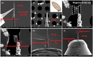
Electron tomography (ET) has gained increasing attention for the 3D characterization of nanoparticles. However, the missing wedge problem due to a limited tilt angle range is still the main challenge for accurate reconstruction in most experimental TEM setups. Advanced algorithms could in-paint or compensate to some extent the missing wedge artifacts, but cannot recover the missing structural information completely. 360° ET provides an option to solve this problem by tilting a needle-shaped specimen over the full tilt range and thus filling the missing information. However, sample preparation especially for fine powders to perform full-range ET is still challenging, thus limiting its application. In this work, we propose a new universal sample preparation method that enables the transfer of selected individual nanoparticle or a few separated nanoparticles by cutting a piece of carbon film supporting the specimen particles and mounting them onto the full-range tomography holder tip with the help of an easily prepared sharp tungsten tip. This method is demonstrated by 360° ET of Pt@TiO2 hollow cage catalyst showing high quality reconstruction without missing wedge.

Type
Software and Instrumentation
Copyright
Copyright © The Author(s), 2022. Published by Cambridge University Press on behalf of the Microscopy Society of America

Access options

Get access to the full version of this content by using one of the access options below. (Log in options will check for institutional or personal access. Content may require purchase if you do not have access.)

References

Apeleo Zubiri, B, Wirth, J, Drobek, D, Englisch, S, Przybilla, T, Weissenberger, T, Schwieger, W & Spiecker, E (2020). Correlative laboratory nano-CT and 360° electron tomography of macropore structures in hierarchical zeolites. Adv Mater Interfaces 8(4), 2001154.CrossRefGoogle Scholar
Arslan, I, Tong, JR & Midgley, PA (2006). Reducing the missing wedge: High-resolution dual axis tomography of inorganic materials. Ultramicroscopy 106(11–12), 9941000.CrossRefGoogle ScholarPubMed
Batenburg, KJ & Sijbers, J (2007). DART: A fast heuristic algebraic reconstruction algorithm for discrete tomography. IEEE.CrossRefGoogle Scholar
Batenburg, KJ & Sijbers, J (2011). DART: A practical reconstruction algorithm for discrete tomography. IEEE Trans Image Process 20(9), 25422553.CrossRefGoogle ScholarPubMed
Biermans, E, Molina, L, Batenburg, KJ, Bals, S & Van Tendeloo, G (2010). Measuring porosity at the nanoscale by quantitative electron tomography. Nano Lett 10(12), 50145019.CrossRefGoogle ScholarPubMed
Bryant, PJ, Kim, HS, Zheng, YC & Yang, R (1987). Technique for shaping scanning tunneling microscope tips. Rev Sci Instrum 58(6), 11151115.CrossRefGoogle Scholar
Distaso, M, Apeleo Zubiri, B, Mohtasebi, A, Inayat, A, Dudák, M, Kočí, P, Butz, B, Klupp Taylor, R, Schwieger, W, Spiecker, E & Peukert, W (2017). Three-dimensional and quantitative reconstruction of non-accessible internal porosity in hematite nanoreactors using 360° electron tomography. Microporous Mesoporous Mater 246, 207214.CrossRefGoogle Scholar
Donoho, DL (2006). Compressed sensing. IEEE Trans Inf Theory 52(4), 12891306.CrossRefGoogle Scholar
Goris, B, Van den Broek, W, Batenburg, KJ, Heidari Mezerji, H & Bals, S (2012). Electron tomography based on a total variation minimization reconstruction technique. Ultramicroscopy 113, 120130.CrossRefGoogle Scholar
Gorji, S, Kashiwar, A, Mantha, LS, Kruk, R, Witte, R, Marek, P, Hahn, H, Kubel, C & Scherer, T (2020). Nanowire facilitated transfer of sensitive TEM samples in a FIB. Ultramicroscopy 219, 113075.CrossRefGoogle ScholarPubMed
Huang, X, Xia, Y, Cao, Y, Zheng, X, Pan, H, Zhu, J, Ma, C, Wang, H, Li, J, You, R, Wei, S, Huang, W & Lu, J (2017). Enhancing both selectivity and coking-resistance of a single-atom Pd1/C3N4 catalyst for acetylene hydrogenation. Nano Res 10(4), 13021312.CrossRefGoogle Scholar
Ibe, JP, Bey, PP Jr, Brandow, SL, Brizzolara, RA, Burnham, NA, DiLella, DP, Lee, KP, Marrian, CR & Colton, RJ (1990). On the electrochemical etching of tips for scanning tunneling microscopy. J Vac Sci Technol, A 8(4), 35703575.CrossRefGoogle Scholar
Jarausch, K & Leonard, DN (2009). Three-dimensional electron microscopy of individual nanoparticles. J Electron Microsc (Tokyo) 58(3), 175183.CrossRefGoogle ScholarPubMed
Kawase, N, Kato, M, Nishioka, H & Jinnai, H (2007). Transmission electron microtomography without the “missing wedge” for quantitative structural analysis. Ultramicroscopy 107(1), 815.CrossRefGoogle ScholarPubMed
Ke, X, Bals, S, Cott, D, Hantschel, T, Bender, H & Van Tendeloo, G (2010). Three-dimensional analysis of carbon nanotube networks in interconnects by electron tomography without missing wedge artifacts. Microsc Microanal 16(2), 210217.CrossRefGoogle ScholarPubMed
Khan, Y, Al-Falih, H, Zhang, Y, Ng, TK & Ooi, BS (2012). Two-step controllable electrochemical etching of tungsten scanning probe microscopy tips. Rev Sci Instrum 83(6), 063708.CrossRefGoogle ScholarPubMed
Kim, CK, Lee, G-J, Lee, MK & Rhee, CK (2014). A novel method to prepare Cu@Ag core–shell nanoparticles for printed flexible electronics. Powder Technol 263, 16.CrossRefGoogle Scholar
Kremer, JR, Mastronarde, DN & McIntosh, JR (1996). Computer visualization of three-dimensional image data using IMOD. J Struct Biol 116, 7176.CrossRefGoogle ScholarPubMed
Kupsch, A, Lange, A, Hentschel, MP, Luck, S, Schmidt, V, Grothausmann, R, Hilger, A & Manke, I (2015). Missing wedge computed tomography by iterative algorithm DIRECTT. J Microsc 261(1), 3645.CrossRefGoogle ScholarPubMed
Lange, A, Kupsch, A, Hentschel, MP, Manke, I, Kardjilov, N, Arlt, T & Grothausmann, R (2011). Reconstruction of limited computed tomography data of fuel cell components using direct iterative reconstruction of computed tomography trajectories. J Power Sources 196(12), 52935298.CrossRefGoogle Scholar
Lanzavecchia, S, Cantele, F, Bellon, PL, Zampighi, L, Kreman, M, Wright, E & Zampighi, GA (2005). Conical tomography of freeze-fracture replicas: A method for the study of integral membrane proteins inserted in phospholipid bilayers. J Struct Biol 149(1), 8798.CrossRefGoogle Scholar
Larson, DJ, Prosa, T, Ulfig, RM, Geiser, BP & Kelly, TF (2013). Local Electrode Atom Probe Tomography, vol. 2. New York, USA: Springer Science.CrossRefGoogle Scholar
Liebertseder, M, Wang, D, Cavusoglu, G, Casapu, M, Wang, S, Behrens, S, Kubel, C, Grunwaldt, JD & Feldmann, C (2021). NaCl-template-based synthesis of TiO2-Pd/Pt hollow nanospheres for H2O2 direct synthesis and CO oxidation. Nanoscale 13(3), 20052011.CrossRefGoogle ScholarPubMed
Mastronarde, DN (1997). Dual-axis tomography: An approach with alignment methods that preserve resolution. J Struct Biol 120(3), 343352.CrossRefGoogle ScholarPubMed
Miao, J, Ercius, P & Billinge, SJ (2016). Atomic electron tomography: 3D structures without crystals. Science 353(6306), aaf2157.CrossRefGoogle ScholarPubMed
Mohanraj, VJ & Chen, Y (2006). Nanoparticles – A review. Trop J Pharm Res 5(1), 561573.Google Scholar
Padgett, E, Hovden, R, DaSilva, JC, Levin, BDA, Grazul, JL, Hanrath, T & Muller, DA (2017). A simple preparation method for full-range electron tomography of nanoparticles and fine powders. Microsc Microanal 23(6), 11501158.CrossRefGoogle ScholarPubMed
Patane, S, Cefali, E, Arena, A, Gucciardi, PG & Allegrini, M (2006). Wide angle near-field optical probes by reverse tube etching. Ultramicroscopy 106(6), 475479.CrossRefGoogle ScholarPubMed
Perezjuste, J, Pastorizasantos, I, Lizmarzan, L & Mulvaney, P (2005). Gold nanorods: Synthesis, characterization and applications. Coord Chem Rev 249(17-18), 18701901.CrossRefGoogle Scholar
Przybilla, T, Zubiri, BA, Beltrán, AM, Butz, B, Machoke, AGF, Inayat, A, Distaso, M, Peukert, W, Schwieger, W & Spiecker, E (2018). Transfer of individual micro- and nanoparticles for high-precision 3D analysis using 360° electron tomography. Small Methods 2(1), 1077276.Google Scholar
Scott, MC, Chen, CC, Mecklenburg, M, Zhu, C, Xu, R, Ercius, P, Dahmen, U, Regan, BC & Miao, J (2012). Electron tomography at 2.4-angstrom resolution. Nature 483(7390), 444447.CrossRefGoogle ScholarPubMed
Sriram, M, Zong, K, Vivekchand, SR & Gooding, JJ (2015). Single nanoparticle plasmonic sensors. Sensors (Basel) 15(10), 2577425792.CrossRefGoogle ScholarPubMed
Tao, AR, Habas, S & Yang, P (2008). Shape control of colloidal metal nanocrystals. Small 4(3), 310325.CrossRefGoogle Scholar
Tofighi, G, Gaur, A, Doronkin, DE, Lichtenberg, H, Wang, W, Wang, D, Rinke, G, Ewinger, A, Dittmeyer, R & Grunwaldt, J-D (2018). Microfluidic synthesis of ultrasmall AuPd nanoparticles with a homogeneously mixed alloy structure in fast continuous flow for catalytic applications. J Phys Chem C 122(3), 17211731.CrossRefGoogle Scholar
Trampert, P, Wang, W, Chen, D, Ravelli, RBG, Dahmen, T, Peters, PJ, Kubel, C & Slusallek, P (2018). Exemplar-based inpainting as a solution to the missing wedge problem in electron tomography. Ultramicroscopy 191, 110.CrossRefGoogle Scholar
Wang, W, Svidrytski, A, Wang, D, Villa, A, Hahn, H, Tallarek, U & Kubel, C (2019). Quantifying morphology and diffusion properties of mesoporous carbon from high-fidelity 3D reconstructions. Microsc Microanal 25(4), 891902.CrossRefGoogle ScholarPubMed
Xu, R, Chen, CC, Wu, L, Scott, MC, Theis, W, Ophus, C, Bartels, M, Yang, Y, Ramezani-Dakhel, H, Sawaya, MR, Heinz, H, Marks, LD, Ercius, P & Miao, J (2015). Three-dimensional coordinates of individual atoms in materials revealed by electron tomography. Nat Mater 14(11), 10991103.CrossRefGoogle ScholarPubMed
Supplementary material: File

Huang et al. supplementary material

Figures S1-S8

Download Huang et al. supplementary material(File)
File 4.3 MB