Hostname: page-component-78c5997874-v9fdk Total loading time: 0 Render date: 2024-11-19T12:33:08.848Z Has data issue: false hasContentIssue false

Optimal STEM Convergence Angle Selection Using a Convolutional Neural Network and the Strehl Ratio

Published online by Cambridge University Press:  06 August 2020
Noah Schnitzer
Affiliation:
Department of Materials Science and Engineering, University of Michigan, Ann Arbor, MI48019, USA Department of Materials Science and Engineering, Cornell University, Ithaca, NY14853, USA
Suk Hyun Sung
Affiliation:
Department of Materials Science and Engineering, University of Michigan, Ann Arbor, MI48019, USA
Robert Hovden*
Affiliation:
Department of Materials Science and Engineering, University of Michigan, Ann Arbor, MI48019, USA Applied Physics Program, University of Michigan, Ann Arbor, MI48109, USA
*
*Author for correspondence: Robert Hovden, E-mail: [email protected]
Get access

Abstract

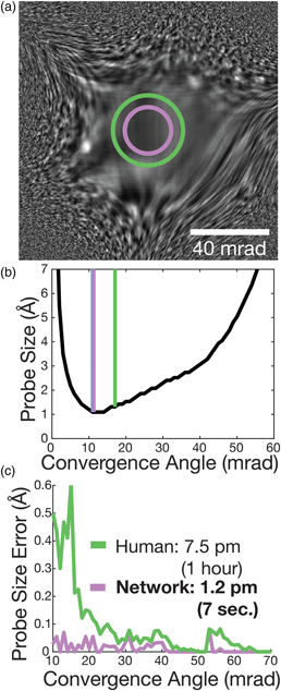
The selection of the correct convergence angle is essential for achieving the highest resolution imaging in scanning transmission electron microscopy (STEM). The use of poor heuristics, such as Rayleigh's quarter-phase rule, to assess probe quality and uncertainties in the measurement of the aberration function results in the incorrect selection of convergence angles and lower resolution. Here, we show that the Strehl ratio provides an accurate and efficient way to calculate criteria for evaluating the probe size for STEM. A convolutional neural network trained on the Strehl ratio is shown to outperform experienced microscopists at selecting a convergence angle from a single electron Ronchigram using simulated datasets. Generating tens of thousands of simulated Ronchigram examples, the network is trained to select convergence angles yielding probes on average 85% nearer to optimal size at millisecond speeds (0.02% of human assessment time). Qualitative assessment on experimental Ronchigrams with intentionally introduced aberrations suggests that trends in the optimal convergence angle size are well modeled but high accuracy requires a high number of training datasets. This near-immediate assessment of Ronchigrams using the Strehl ratio and machine learning highlights a viable path toward the rapid, automated alignment of aberration-corrected electron microscopes.

Type
Software and Instrumentation
Copyright
Copyright © Microscopy Society of America 2020

Access options

Get access to the full version of this content by using one of the access options below. (Log in options will check for institutional or personal access. Content may require purchase if you do not have access.)

References

Badrinarayanan, V, Kendall, A & Cipolla, R (2017). SegNet: A deep convolutional encoder-decoder architecture for image segmentation. IEEE Trans Pattern Anal Mach Intell 39, 24812495.CrossRefGoogle ScholarPubMed
Barthel, J (2018). Dr. Probe: A software for high-resolution STEM image simulation. Ultramicroscopy 193, 111.CrossRefGoogle ScholarPubMed
Burke, D, Patton, B, Huang, F, Bewersdorf, J & Booth, MJ (2015). Adaptive optics correction of specimen-induced aberrations in single-molecule switching microscopy. Optica 2, 177185.CrossRefGoogle Scholar
Cowley, JM (1979 a). Coherent interference in convergent-beam electron diffraction and shadow imaging. Ultramicroscopy 4, 435449.CrossRefGoogle Scholar
Cowley, JM (1979 b). Adjustment of a STEM instrument by use of shadow images. Ultramicroscopy 4, 413418.CrossRefGoogle Scholar
Cowley, JM (1980). Interference effects in a stem instrument. Micron (1969) 11, 229233.CrossRefGoogle Scholar
Crewe, AV & Salzman, DB (1982). On the optimum resolution for a corrected STEM. Ultramicroscopy 9, 373378.CrossRefGoogle Scholar
Dellby, N, Krivanek, L, Nellist, D, Batson, E & Lupini, R (2001). Progress in aberration-corrected scanning transmission electron microscopy. Microscopy 50, 177185.CrossRefGoogle ScholarPubMed
Deng, J, Dong, W, Socher, R, Li, L, Li, K & Fei-Fei, L (2009). ImageNet: A large-scale hierarchical image database. In 2009 IEEE Conference on Computer Vision and Pattern Recognition, Miami, FL, 20-25 July 2009, pp. 248–255.CrossRefGoogle Scholar
Dwyer, C, Erni, R & Etheridge, J (2010). Measurement of effective source distribution and its importance for quantitative interpretation of STEM images. Ultramicroscopy 110, 952957.CrossRefGoogle Scholar
Ede, JM & Beanland, R (2019). Improving electron micrograph signal-to-noise with an atrous convolutional encoder-decoder. Ultramicroscopy 202, 1825.CrossRefGoogle ScholarPubMed
Haider, M, Uhlemann, S & Zach, J (2000). Upper limits for the residual aberrations of a high-resolution aberration-corrected STEM. Ultramicroscopy 81, 163175.CrossRefGoogle ScholarPubMed
Henstra, A & Tiemeijer, PC (2018). Aberration measurement in a charged particle microscope. Available at https://patents.google.com/patent/US10157727B2/en?q=ceos+corrector&oq=ceos+corrector (accessed April 6, 2020).Google Scholar
Janssen, BJ, van Duinen, G, Luecken, U, Savage, R, van den Boom, SHL & Lazic, I (2015). Method of investigating the wavefront of a charged-particle beam. Available at https://patents.google.com/patent/US20150170876A1/en?q=ceos+corrector&oq=ceos+corrector (accessed April 6, 2020).Google Scholar
Kirkland, EJ (2010). Advanced Computing in Electron Microscopy, 2nd ed. Springer, USA. Available at https://www.springer.com/us/book/9781441965325 (accessed September 4, 2018).CrossRefGoogle Scholar
Kirkland, EJ (2011). On the optimum probe in aberration corrected ADF-STEM. Ultramicroscopy 111, 15231530.CrossRefGoogle ScholarPubMed
Kirkland, EJ (2018). Fine tuning an aberration corrected ADF-STEM. Ultramicroscopy 186, 6265.CrossRefGoogle ScholarPubMed
Krivanek, OL, Dellby, N, Keyse, RJ, Murfitt, MF, Own, CS & Szilagyi, ZS (2008). Advances in aberration-corrected scanning transmission electron microscopy and electron energy-loss spectroscopy. In Advances in Imaging and Electron Physics, vol. 153, Hawkes, PW (Ed.), pp. 121160.San Diego, CA: Academic Press.Google Scholar
Krivanek, OL, Dellby, N & Lupini, AR (1999). Towards sub-Å electron beams. Ultramicroscopy 78, 111.CrossRefGoogle Scholar
Krivanek, OL, Dellby, N, Spence, AJ, Camps, RA & Brown, LM (1997). Aberration correction in the STEM. In Institute of Physics Conference Series 153 (Proceedings 1997 EMAG Meeting), Rodenburg JM (Ed.), pp. 35–40. Bristol, UK: Institute of Physics Publishing.Google Scholar
Krizhevsky, A, Sutskever, I & Hinton, GE (2012). ImageNet classification with deep convolutional neural networks. In Advances in Neural Information Processing Systems, vol. 25, Pereira, F, Burges, CJC, Bottou, L & Weinberger, KQ (Eds.), pp. 10971105. Curran Associates, Inc. Available at http://papers.nips.cc/paper/4824-imagenet-classification-with-deep-convolutional-neural-networks.pdf (accessed July 30, 2019).Google Scholar
Lathuiliére, S, Mesejo, P, Alameda-Pineda, X & Horaud, R (2019). A comprehensive analysis of deep regression. IEEE Trans Pattern Anal Mach Intell, 11.Google ScholarPubMed
Lazic, I, van Duinen, G & Tiemeijer, PC (2014). Method of investigating and correcting aberrations in a charged-particle lens system. Available at https://patents.google.com/patent/US20140061464A1/en?q=ceos+corrector&oq=ceos+corrector (accessed April 6, 2020).Google Scholar
LeCun, Y, Bengio, Y & Hinton, G (2015). Deep learning. Nature 521, 436444.CrossRefGoogle ScholarPubMed
LeCun, Y, Bottou, L, Bengio, Y & Haffner, P (1998). Gradient-based learning applied to document recognition. Proc IEEE 86, 22782324.CrossRefGoogle Scholar
Lin, JA & Cowley, JM (1986). Calibration of the operating parameters for an HB5 stem instrument. Ultramicroscopy 19, 3142.CrossRefGoogle Scholar
Lupini, AR, Wang, P, Nellist, PD, Kirkland, AI & Pennycook, SJ (2010). Aberration measurement using the Ronchigram constrast transfer function. Ultramicroscopy 110, 891898.CrossRefGoogle Scholar
Mahajan, VN (1982). Strehl ratio for primary aberrations: Some analytical results for circular and annular pupils. JOSA 72, 12581266.CrossRefGoogle Scholar
Mahendran, S, Ali, H & Vidal, R (2017). 3D pose regression using convolutional neural networks. In 2017 IEEE International Conference on Computer Vision Workshops (ICCVW), Venice, Italy, Oct. 22-29 2017, pp. 2174–2182.Google Scholar
Müller, H, Uhlemann, S, Hartel, P & Haider, M (2006). Advancing the hexapole Cs-corrector for the scanning transmission electron microscope. Microsc Microanal 12, 442455.CrossRefGoogle ScholarPubMed
Ophus, C (2017). A fast image simulation algorithm for scanning transmission electron microscopy. Adv Struct Chem Imaging 3, 13.CrossRefGoogle ScholarPubMed
Porro, IL, Traub, WA & Carleton, NP (1999). Effect of telescope alignment on a stellar interferometer. Appl Optics 38, 60556067.CrossRefGoogle ScholarPubMed
Rayleigh, L (1879). XXXI. Investigations in optics, with special reference to the spectroscope. London, Edinburgh Dublin Philos Mag J Sci 8, 261274.CrossRefGoogle Scholar
Rayleigh, L (1896). XV. On the theory of optical images, with special reference to the microscope. London, Edinburgh Dublin Philos Mag J Sci 42, 167195.CrossRefGoogle Scholar
Ronchi, V (1923). Due nuovi metodi per lo studio delle superficie e dei sistemi ottici. Il Nuovo Cimento 26, 6971.CrossRefGoogle Scholar
Ronchi, V (1964). Forty years of history of a grating interferometer. Appl Optics 3, 437451.CrossRefGoogle Scholar
Rose, H (1981). Correction of aperture aberrations in magnetic systems with threefold symmetry. Nucl Instrum Methods Phys Res 187, 187199.CrossRefGoogle Scholar
Sasaki, T, Sawada, H, Hosokawa, F, Kohno, Y, Tomita, T, Kaneyama, T, Kondo, Y, Kimoto, K, Sato, Y & Suenaga, K (2010). Performance of low-voltage STEM/TEM with delta corrector and cold field emission gun. J Electron Microsc 59, S7S13.CrossRefGoogle ScholarPubMed
Sawada, H, Sannomiya, T, Hosokawa, F, Nakamichi, T, Kaneyama, T, Tomita, T, Kondo, Y, Tanaka, T, Oshima, Y, Tanishiro, Y & Takayanagi, K (2008). Measurement method of aberration from Ronchigram by autocorrelation function. Ultramicroscopy 108, 14671475.CrossRefGoogle ScholarPubMed
Scherzer, O (1949). The theoretical resolution limit of the electron microscope. J Appl Phys 20, 2029.CrossRefGoogle Scholar
Schnitzer, N, Sung, SH & Hovden, R (2019). Introduction to the Ronchigram and its calculation with Ronchigram.com. Microsc Today 27, 1215.CrossRefGoogle Scholar
Weyland, M & Muller, DA (2005). Tuning the convergence angle for optimum STEM performance. FEI NanoSolutions 1, 2435.Google Scholar
Wong, K, Kirkland, E, Xu, P, Loane, R & Silcox, J (1992). Measurement of spherical aberration in STEM. Ultramicroscopy 40, 139150.CrossRefGoogle Scholar
Xu, W & LeBeau, JM (2018). A deep convolutional neural network to analyze position averaged convergent beam electron diffraction patterns. Ultramicroscopy 188, 5969.CrossRefGoogle ScholarPubMed
Zhang, C, Feng, J, DaCosta, LR & Voyles, PM (2020). Atomic resolution convergent beam electron diffraction analysis using convolutional neural networks. Ultramicroscopy 210, 112921.CrossRefGoogle Scholar
Supplementary material: File

Schnitzer et al. supplementary material

Tables S1-S2 and Figures S1-S2

Download Schnitzer et al. supplementary material(File)
File 156.2 KB