Hostname: page-component-586b7cd67f-r5fsc Total loading time: 0 Render date: 2024-11-26T06:02:18.712Z Has data issue: false hasContentIssue false

Nanoscale Visualization of Phase Transition in Melting of Sn–Bi Particles by In situ Hard X-ray Ptychographic Coherent Diffraction Imaging

Published online by Cambridge University Press:  28 August 2020
Nozomu Ishiguro
Affiliation:
Tohoku University, Institute of Multidisciplinary Research for Advanced Materials (IMRAM)2-1-1, Katahira, Aoba-ku, Sendai980-0857, Miyagi, Japan RIKEN SPring-8 Center, 1-1-1, Kouto, Sayo-cho, Sayo, Hyogo679-5148, Japan
Takaya Higashino
Affiliation:
RIKEN SPring-8 Center, 1-1-1, Kouto, Sayo-cho, Sayo, Hyogo679-5148, Japan Graduate School of Engineering, Osaka University, 2-1 Yamada-oka, Suita, Osaka565-0871, Japan
Makoto Hirose
Affiliation:
RIKEN SPring-8 Center, 1-1-1, Kouto, Sayo-cho, Sayo, Hyogo679-5148, Japan Graduate School of Engineering, Osaka University, 2-1 Yamada-oka, Suita, Osaka565-0871, Japan
Yukio Takahashi*
Affiliation:
Tohoku University, Institute of Multidisciplinary Research for Advanced Materials (IMRAM)2-1-1, Katahira, Aoba-ku, Sendai980-0857, Miyagi, Japan RIKEN SPring-8 Center, 1-1-1, Kouto, Sayo-cho, Sayo, Hyogo679-5148, Japan Graduate School of Engineering, Osaka University, 2-1 Yamada-oka, Suita, Osaka565-0871, Japan Tohoku University, International Center for Synchrotron Radiation Innovation Smart (SRIS)2-1-1, Katahira, Aoba-ku, Sendai980-0857, Miyagi, Japan
*
*Author for correspondence: Yukio Takahashi, E-mail: [email protected]
Get access

Abstract

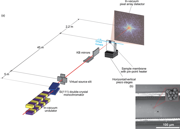
The phase transition in the melting of SnBi eutectic solder alloy particles was observed by in situ hard X-ray ptychographic coherent diffraction imaging with a pin-point heating system. Ptychographic diffraction patterns of micrometer-sized SnBi particles were collected at temperatures from room temperature to 540 K. The projection images of each particle were reconstructed at a spatial resolution of 25 nm, showing differences in the phase shifts due to two crystal phases in the SnBi alloy system and the Sn/Bi oxides at the surface. By quantitatively evaluating the Bi content, it became clear that the nonuniformity of the composition of Sn and Bi at the single-particle level exists when the particles are synthesized by centrifugal atomization.

Type
Materials Science Applications
Copyright
Copyright © Microscopy Society of America 2020

Access options

Get access to the full version of this content by using one of the access options below. (Log in options will check for institutional or personal access. Content may require purchase if you do not have access.)

Footnotes

a

Nozomu Ishiguro and Takaya Higashino contributed equally to this work.

References

Alessandro, FS, Walman, BC, Benedito, AL & Claudio, SK (2004). Microstructure of under-cooled Sn–Bi and Al–Si alloys. Mater Sci Eng A 375–377, 473478.Google Scholar
Bhattacharya, V & Chattopadhyay, K (2007). Melting of multiphase nano-scaled particles embedded in Al matrix: Case of Pb–Sn and Bi–Sn alloys. Mater Sci Eng A 449–451, 10031008.CrossRefGoogle Scholar
Buurmans, ILC & Weckhuysen, BM (2012). Heterogeneities of individual catalyst particles in space and time as monitored by spectroscopy. Nat Chem 4, 873886.CrossRefGoogle ScholarPubMed
Chapman, HN, Barty, A, Marchesini, S, Noy, A & Hau-Riege, SP (2006). High-resolution ab initio three-dimensional X-ray diffraction microscopy. J Opt Soc Am A 23, 11791200.CrossRefGoogle ScholarPubMed
Chapman, HN & Nugent, KA (2010). Coherent lensless X-ray imaging. Nat Photonics 4, 833839.CrossRefGoogle Scholar
Creemer, JF, Helveg, S, Hoveling, GH, Ullmann, S, Molenbroek, AM, Sarro, PM & Zandbergen, HW (2008). Atomic-scale electron microscopy at ambient pressure. Ultramicroscopy 108, 993998.CrossRefGoogle ScholarPubMed
Davis, TJ (1994). A unified treatment of small-angle X-ray scattering, X-ray refraction and absorption using the Rytov approximation. Acta Cryst A 50, 686690.CrossRefGoogle Scholar
Henke, BL, Gullikson, EM & Davis, JC (1993). X-ray interactions: Photo absorption, scattering, transmission, and reflection at E=50-30000 eV, Z=1–92. At Data Nucl Data Tables 54, 181342.CrossRefGoogle Scholar
Høydalsvik, K, Fløystad, JB, Zhao, T, Esmaeili, M, Diaz, A, Andreasen, JW, Mathiesen, RH, Rønning, M & Breiby, DW (2014). In situ X-ray ptychography imaging of high-temperature CO2 acceptor particle agglomerates. Appl Phys Lett 104, 241909.CrossRefGoogle Scholar
Maiden, AM & Rodenburg, JM (2009). An improved ptychographical phase retrieval algorithm for diffractive imaging. Ultramicroscopy 109, 12561262.CrossRefGoogle ScholarPubMed
Miao, J, Charalambous, P, Kirz, J & Sayre, D (1999). Extending the methodology of X-ray crystallography to allow imaging of micrometre-sized non-crystalline specimens. Nature 400, 342344.CrossRefGoogle Scholar
Pfeiffer, F (2017). X-ray ptychography. Nat Photonics 12, 917.CrossRefGoogle Scholar
Robinson, I & Harder, R (2009). Coherent X-ray diffraction imaging of strain at the nanoscale. Nat Mater 8, 219298.CrossRefGoogle ScholarPubMed
Rodenburg, JM, Hurst, AC, Cullis, AG, Dobson, BR, Pfeiffer, F, Bunk, O, David, C, Jefimovs, K & Johnson, I (2007). Hard-X-ray lensless imaging of extended objects. Phys Rev Lett 98, 034801.CrossRefGoogle ScholarPubMed
Sakdinawat, A & Attwood, D (2010). Nanoscale X-ray imaging. Nat Photonics 4, 840848.CrossRefGoogle Scholar
Shao, M (2014). In situ microscopic studies on the structural and chemical behaviors of lithium-ion battery materials. J Power Sources 270, 475486.CrossRefGoogle Scholar
Suzana, AF, Rochet, A, Passos, AR, Zerba, JPC, Polo, CC, Santilli, CV, Pulcinelli, SH, Berenguer, F, Harder, R, Maxeyd, E & Meneau, F (2019). In situ three-dimensional imaging of strain in gold nanocrystals during catalytic oxidation. Nanoscale Adv 1, 30093014.CrossRefGoogle Scholar
Takahashi, Y, Suzuki, A, Zettsu, N, Kohmura, Y, Senba, Y, Ohashi, H, Yamauchi, K & Ishikawa, T (2011). Towards high-resolution ptychographic X-ray diffraction microscopy. Phys Rev B 83, 214109.CrossRefGoogle Scholar
Ulvestad, A, Singer, A, Clark, JN, Cho, HM, Kim, JW, Harder, R, Maser, J, Meng, YS & Shpyrko, OG (2015). Topological defect dynamics in operando battery nanoparticles. Science 348, 13441347.CrossRefGoogle ScholarPubMed
Warren, BE (1990). X-ray Diffraction. New York, NY: Dover Publications.Google Scholar