Hostname: page-component-586b7cd67f-t7fkt Total loading time: 0 Render date: 2024-11-23T06:54:16.444Z Has data issue: false hasContentIssue false

Multi-modal characterization of collagen fibril orientation in human cortical bone by a combination of quantitative polarized Raman spectroscopy, nanoscale X-ray computed tomography and 360° electron tomography

Published online by Cambridge University Press:  30 July 2021

Tatiana Kormilina
Affiliation:
Institute of Micro- and Nanostructure Research (IMN) & Center for Nanoanalysis and Electron Microscopy (CENEM), Interdisciplinary Center for Nanostructured Films (IZNF), Department of Materials Science and Engineering, Friedrich-Alexander University Erlangen-Nürnberg (FAU), Erlangen, Germany, United States
Silvan Englisch
Affiliation:
Institute of Micro- and Nanostructure Research (IMN) & Center for Nanoanalysis and Electron Microscopy (CENEM), Interdisciplinary Center for Nanostructured Films (IZNF), Department of Materials Science and Engineering, Friedrich-Alexander University Erlangen-Nürnberg (FAU), Erlangen, Germany, Erlangen, Bayern, Germany
Tatiana Kochetkova
Affiliation:
Empa, Swiss Federal Laboratories for Materials Science and Technology, Laboratory for Mechanics of Materials & Nanostructures, Thun, Switzerland, United States
Dominik Drobek
Affiliation:
Institute of Micro- and Nanostructure Research (IMN) & Center for Nanoanalysis and Electron Microscopy (CENEM), Interdisciplinary Center for Nanostructured Films (IZNF), Department of Materials Science and Engineering, Friedrich-Alexander University Erlangen-Nürnberg (FAU), Erlangen, Germany, United States
Janis Wirth
Affiliation:
Institute of Micro- and Nanostructure Research (IMN) & Center for Nanoanalysis and Electron Microscopy (CENEM), Interdisciplinary Center for Nanostructured Films (IZNF), Department of Materials Science and Engineering, Friedrich-Alexander University Erlangen-Nürnberg (FAU), Erlangen, Germany, Erlangen, Bayern, Germany
Benjamin Apeleo Zubiri
Affiliation:
Institute of Micro- and Nanostructure Research (IMN) & Center for Nanoanalysis and Electron Microscopy (CENEM), Interdisciplinary Center for Nanostructured Films (IZNF), Department of Materials Science and Engineering, Friedrich-Alexander University Erlangen-Nürnberg (FAU), Erlangen, Germany, Erlangen, Bayern, Germany
Jakob Schwiedrzik
Affiliation:
Empa, Swiss Federal Laboratories for Materials Science and Technology, Laboratory for Mechanics of Materials & Nanostructures, Thun, Switzerland, United States
Erdmann Spiecker
Affiliation:
Institute of Micro- and Nanostructure Research (IMN) & Center for Nanoanalysis and Electron Microscopy (CENEM), Interdisciplinary Center for Nanostructured Films (IZNF), Department of Materials Science and Engineering, Friedrich-Alexander University Erlangen-Nürnberg (FAU), Erlangen, Germany, Erlangen, Bayern, Germany

Abstract

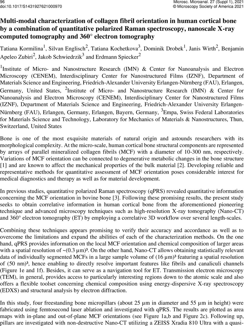
Image of the first page of this content. For PDF version, please use the ‘Save PDF’ preceeding this image.'
Type
Multi-Modal Multi-Dimensional Microscopy
Copyright
Copyright © The Author(s), 2021. Published by Cambridge University Press on behalf of the Microscopy Society of America

References

Acerbo, A. S., et al. "Alterations in collagen and mineral nanostructure observed in osteoporosis and pharmaceutical treatments using simultaneous small-and wide-angle X-ray scattering." Calcified tissue international 95.5 (2014): 446-456.CrossRefGoogle ScholarPubMed
Zimmermann, E. A., et al. "Intrinsic mechanical behavior of femoral cortical bone in young, osteoporotic and bisphosphonate-treated individuals in low-and high energy fracture conditions." Scientific reports 6.1 (2016): 1-12.Google ScholarPubMed
Kochetkova, T., et al. "Combining polarized Raman spectroscopy and micropillar compression to study microscale structure-property relationships in mineralized tissues." Acta biomaterialia 119 (2021): 390-404.CrossRefGoogle ScholarPubMed
Sommer, C., et al. "Ilastik: Interactive learning and segmentation toolkit." 2011 IEEE international symposium on biomedical imaging: From nano to macro. IEEE, 2011.Google Scholar
We gratefully acknowledge financial support by the German Research Foundation (DFG) within the frameworks of the research training group GRK1896 “In situ Microscopy with Electrons, X-rays and Scanning Probes”, the project SP648/8 “High-resolution X-ray microscopy for correlative tomography, high throughput screening and in situ mechanical testing of structural and functional materials” (Project-ID 316992193), the Collaborative Research Centre 1411 “Design of Particulate Products” (Project-ID 416229255) and the Collaborative Research Centre 1452 "Catalysis at Liquid Interfaces", as well as Special Focus Area Personalized Health and Related Technologies (SFA PHRT) iDoc Project 2017-304 and Swiss National Science Foundation (SNSF) Ambizione grant no. 174192.Google Scholar