Hostname: page-component-586b7cd67f-2plfb Total loading time: 0 Render date: 2024-11-25T23:06:11.288Z Has data issue: false hasContentIssue false

Laboratory Soft X-Ray Microscopy with an Integrated Visible-Light Microscope—Correlative Workflow for Faster 3D Cell Imaging

Published online by Cambridge University Press:  07 October 2020

Aurélie Dehlinger*
Affiliation:
Technische Universität Berlin, Institut für Optik und Atomare Physik, Hardenbergstraße 36, Berlin 10623, Germany Berlin Laboratory for Innovative X-ray technologies (BLiX), Hardenbergstraße 36, Berlin 10623, Germany
Christian Seim
Affiliation:
Technische Universität Berlin, Institut für Optik und Atomare Physik, Hardenbergstraße 36, Berlin 10623, Germany Berlin Laboratory for Innovative X-ray technologies (BLiX), Hardenbergstraße 36, Berlin 10623, Germany
Holger Stiel
Affiliation:
Berlin Laboratory for Innovative X-ray technologies (BLiX), Hardenbergstraße 36, Berlin 10623, Germany Max-Born-Institut (MBI) im Forschungsverbund Berlin e.V., Max-Born-Straße 2A, Berlin 12489, Germany
Shailey Twamley
Affiliation:
Charité – Universitätsmedizin Berlin, corporate member of Freie Universität Berlin, Humboldt-Universität zu Berlin, and Berlin Institute of Health, Medizinische Klinik für Kardiologie und Angiologie, Campus Mitte, Charitéplatz 1, 10117 Berlin, Germany DZHK (German Centre for Cardiovascular Research), Partner Site Berlin, Charitéplatz 1, 10117 Berlin, Germany
Antje Ludwig
Affiliation:
Charité – Universitätsmedizin Berlin, corporate member of Freie Universität Berlin, Humboldt-Universität zu Berlin, and Berlin Institute of Health, Medizinische Klinik für Kardiologie und Angiologie, Campus Mitte, Charitéplatz 1, 10117 Berlin, Germany DZHK (German Centre for Cardiovascular Research), Partner Site Berlin, Charitéplatz 1, 10117 Berlin, Germany Charité – Universitätsmedizin Berlin, corporate member of Freie Universität Berlin, Humboldt-Universität zu Berlin, and Berlin Institute of Health, Klinik für Radiologie, Charitéplatz 1, 10117 Berlin, Germany
Mikael Kördel
Affiliation:
Department of Applied Physics, KTH Royal Institute of Technology/Albanova, Stockholm 106 91, Sweden
Daniel Grötzsch
Affiliation:
Technische Universität Berlin, Institut für Optik und Atomare Physik, Hardenbergstraße 36, Berlin 10623, Germany Berlin Laboratory for Innovative X-ray technologies (BLiX), Hardenbergstraße 36, Berlin 10623, Germany
Stefan Rehbein
Affiliation:
Helmholtz Zentrum Berlin für Materialien und Energie GmbH, Wilhelm-Conrad-Röntgen Campus, Albert-Einstein-Str. 15, Berlin 12489, Germany
Birgit Kanngießer
Affiliation:
Technische Universität Berlin, Institut für Optik und Atomare Physik, Hardenbergstraße 36, Berlin 10623, Germany Berlin Laboratory for Innovative X-ray technologies (BLiX), Hardenbergstraße 36, Berlin 10623, Germany
*
*Author for correspondence: Aurélie Dehlinger, E-mail: [email protected]
Get access

Abstract

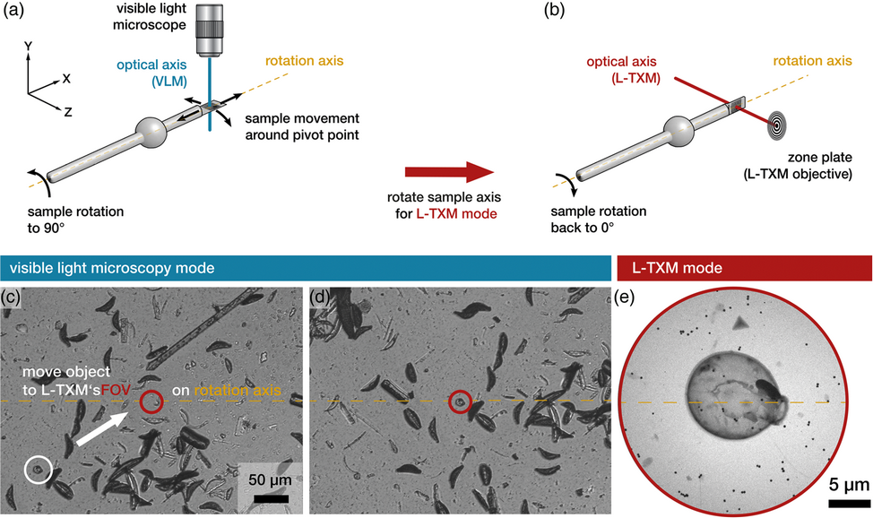
Laboratory transmission soft X-ray microscopy (L-TXM) has emerged as a complementary tool to synchrotron-based TXM and high-resolution biomedical 3D imaging in general in recent years. However, two major operational challenges in L-TXM still need to be addressed: a small field of view and a potentially misaligned rotation stage. As it is not possible to alter the magnification during operation, the field of view in L-TXM is usually limited to a few tens of micrometers. This complicates locating areas and objects of interest in the sample. Additionally, if the rotation axis of the sample stage cannot be adjusted prior to the experiments, an efficient workflow for tomographic imaging cannot be established, as refocusing and sample repositioning will become necessary after each recorded projection. Both these limitations have been overcome with the integration of a visible-light microscope (VLM) into the L-TXM system. Here, we describe the calibration procedure of the goniometer sample stage and the integrated VLM and present the resulting 3D imaging of a test sample. In addition, utilizing this newly integrated VLM, the extracellular matrix of cryofixed THP-1 cells (human acute monocytic leukemia cells) was visualized by L-TXM for the first time in the context of an ongoing biomedical research project.

Type
Software and Instrumentation
Copyright
Copyright © Microscopy Society of America 2020

Access options

Get access to the full version of this content by using one of the access options below. (Log in options will check for institutional or personal access. Content may require purchase if you do not have access.)

References

Born, M & Wolf, E (1999. Chapter 10, Interference and diffraction with partially coherent light. In Principles of Optics, Born, M & Wolf, E (Eds.), pp. 554632. Cambridge: Cambridge University PressCrossRefGoogle Scholar
Chao, W, Fischer, P, Tyliszczak, T, Rekawa, S, Anderson, E & Naulleau, P (2012). Real space soft X-ray imaging at 10 nm spatial resolution. Optics Express 20, 9777.CrossRefGoogle ScholarPubMed
Chiappi, M, Conesa, JJ, Pereiro, E, Sorzano, COS, Rodríguez, MJ, Henzler, K, Schneider, G, Chichón, FJ & Carrascosa, JL (2016). Cryo-soft X-ray tomography as a quantitative three-dimensional tool to model nanoparticle: Cell interaction. J Nanobiotechnol 14, 15. doi:10.1186/s12951-016-0170-4.CrossRefGoogle ScholarPubMed
Cruz-Adalia, A, Ramirez-Santiago, G, Calabia-Linares, C, Torres-Torresano, M, Feo, L, Galán-Díez, M, Fernández-Ruiz, E, Pereiro, E, Guttmann, P, Chiappi, M, Schneider, G, Carrascosa, JL, Chichón, FJ, Martínez Del Hoyo, G, Sánchez-Madrid, F & Veiga, E (2014). T cells kill bacteria captured by transinfection from dendritic cells and confer protection in mice. Cell Host Microbe. 15, 611622.CrossRefGoogle ScholarPubMed
Ebong, EE, MacAluso, FP, Spray, DC & Tarbell, JM (2011). Imaging the endothelial glycocalyx in vitro by rapid freezing/freeze substitution transmission electron microscopy. Arterioscl Thromb Vasc Biol 31, 19081915.CrossRefGoogle ScholarPubMed
Fogelqvist, E, Kördel, M, Carannante, V, Önfelt, B & Hertz, HM (2017). Laboratory cryo X-ray microscopy for 3D cell imaging. Sci Rep 7, 18.CrossRefGoogle ScholarPubMed
Harkiolaki, M, Darrow, MC, Spink, MC, Kosior, E, Dent, K & Duke, E (2018). Cryo-soft X-ray tomography: Using soft X-rays to explore the ultrastructure of whole cells. Emerg Top Life Sci 2, 8192.Google ScholarPubMed
Kapishnikov, S, Staalsø, T, Yang, Y, Lee, J, Pérez-Berná, AJ, Pereiro, E, Yang, Y, Werner, S, Guttmann, P, Leiserowitz, L & Als-Nielsen, J (2019). Mode of action of quinoline antimalarial drugs in red blood cells infected by Plasmodium falciparum revealed in vivo. Proc Natl Acad Sci 116, 2294622952.CrossRefGoogle ScholarPubMed
Kepsutlu, B, Wycisk, V, Achazi, K, Kapishnikov, S, Pérez-Berná, AJ, Guttmann, P, Cossmer, A, Pereiro, E, Ewers, H, Ballauff, M, Schneider, G & McNally, JG (2020). Cells undergo major changes in the quantity of cytoplasmic organelles after uptake of gold nanoparticles with biologically relevant surface coatings. ACS Nano 14, 22482264. doi:10.1021/acsnano.9b09264CrossRefGoogle ScholarPubMed
Kördel, M, Dehlinger, A, Seim, C, Vogt, U, Fogelqvist, E, Sellberg, JA, Stiel, H & Hertz, HM (2020). Laboratory water-window X-ray microscopy. Optica 7, 658674.CrossRefGoogle Scholar
Kremer, JR, Mastronarde, DN & McIntosh, JR (1996). Computer visualization of three-dimensional image data using IMOD. J Struct Biol 116, 7176.CrossRefGoogle ScholarPubMed
Legall, H, Blobel, G, Stiel, H, Sandner, W, Seim, C, Takman, P, Martz, DH, Selin, M, Vogt, U, Hertz, HM, Esser, D, Sipma, H, Luttmann, J, Höfer, M, Hoffmann, HD, Yulin, S, Feigl, T, Rehbein, S, Guttmann, P, Schneider, G, Wiesemann, U, Wirtz, M & Diete, W (2012). Compact X-ray microscope for the water window based on a high brightness laser plasma source. Opt Express 20, 18362.CrossRefGoogle ScholarPubMed
Le Gros, MA, Clowney, EJ, Magklara, A, Yen, A, Markenscoff-Papadimitrou, E, Colquitt, B, Myllys, M, Kellis, M, Lomvardas, S & Larabell, CA (2016). Soft X-ray tomography reveals gradual chromatin compaction and reorganization during neurogenesis in vivo. Cell Rep 17, 21252136.CrossRefGoogle ScholarPubMed
Martz, DH, Selin, M, von Hofsten, O, Fogelqvist, E, Holmberg, A, Vogt, U, Legall, H, Blobel, G, Seim, C, Stiel, H & Hertz, HM (2012). High average brightness water window source for short-exposure cryomicroscopy. Opt Lett 37, 44254427.CrossRefGoogle ScholarPubMed
McDermott, G, Le Gros, MA, Knoechel, CG, Uchida, M & Larabell, CA (2009). Soft X-ray tomography and cryogenic light microscopy: The cool combination in cellular imaging. Trends Cell Biol 19, 587595.CrossRefGoogle ScholarPubMed
Poller, WC, Löwa, N, Schleicher, M, Münster-Wandowski, A, Taupitz, M, Stangl, V, Ludwig, A & Wiekhorst, F (2020). Initial interaction of citrate-coated iron oxide nanoparticles with the glycocalyx of THP-1 monocytes assessed by real-time magnetic particle spectroscopy and electron microscopy. Sci Rep 10, 19.CrossRefGoogle ScholarPubMed
Schneider, G (1998). Cryo X-ray microscopy with high spatial resolution in amplitude and phase contrast. Ultramicroscopy 75, 85104.CrossRefGoogle ScholarPubMed
Schneider, G, Guttmann, P, Rehbein, S, Werner, S & Follath, R (2012). Cryo X-ray microscope with flat sample geometry for correlative fluorescence and nanoscale tomographic imaging. J Struct Biol 177, 212223.CrossRefGoogle ScholarPubMed
Sorrentino, A, Nicolás, J, Valcárcel, R, Chichón, FJ, Rosanes, M, Avila, J, Tkachuk, A, Irwin, J, Ferrer, S & Pereiro, E (2015). MISTRAL: A transmission soft X-ray microscopy beamline for cryo nano-tomography of biological samples and magnetic domains imaging. J Synchrotron Radiat 22, 11121117.CrossRefGoogle ScholarPubMed
Studer, D, Humbel, BM & Chiquet, M (2008). Electron microscopy of high pressure frozen samples: Bridging the gap between cellular ultrastructure and atomic resolution. Histochem Cell Biol 130, 877889.CrossRefGoogle ScholarPubMed