Hostname: page-component-586b7cd67f-l7hp2 Total loading time: 0 Render date: 2024-11-26T07:56:21.492Z Has data issue: false hasContentIssue false

Higher-Order Structure of Human Chromosomes Observed by Electron Diffraction and Electron Tomography

Published online by Cambridge University Press:  20 November 2020
Misa Hayashida*
Affiliation:
Nanotechnology Research Centre, National Research Council of Canada, 11421 Saskatchewan Drive, Edmonton, AB, T6G 2M9, Canada
Rinyaporn Phengchat
Affiliation:
Graduate School of Human Development and Environment, Kobe University, 3-11 Tsurukabuto, Nada-ku, Kobe657-8501, Japan
Marek Malac
Affiliation:
Nanotechnology Research Centre, National Research Council of Canada, 11421 Saskatchewan Drive, Edmonton, AB, T6G 2M9, Canada Department of Physics, University of Alberta, EdmontonT6G 2E1, Canada
Ken Harada
Affiliation:
Center for Emergent Matter Science (CEMS), RIKEN, Hatoyama, Saitama350-0395, Japan
Tetsuya Akashi
Affiliation:
Research & Development Group, HITACHI, Ltd., Hatoyama, Saitama350-0395, Japan
Nobuko Ohmido
Affiliation:
Graduate School of Human Development and Environment, Kobe University, 3-11 Tsurukabuto, Nada-ku, Kobe657-8501, Japan
Kiichi Fukui
Affiliation:
Graduate School of Pharmaceutical Sciences, Osaka University, 1-6 Yamadaoka, Suita, Osaka565-0871, Japan
*
*Author for correspondence: Misa Hayashida, E-mail: [email protected]
Get access

Abstract

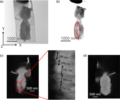
It is well known that two DNA molecules are wrapped around histone octamers and folded together to form a single chromosome. However, the nucleosome fiber folding within a chromosome remains an enigma, and the higher-order structure of chromosomes also is not understood. In this study, we employed electron diffraction which provides a noninvasive analysis to characterize the internal structure of chromosomes. The results revealed the presence of structures with 100–200 nm periodic features directionally perpendicular to the chromosome axis in unlabeled isolated human chromosomes. We also visualized the 100–200 nm periodic features perpendicular to the chromosome axis in an isolated chromosome whose DNA molecules were specifically labeled with OsO4 using electron tomography in 300 keV and 1 MeV transmission electron microscopes.

Type
Biological Applications
Copyright
Copyright © The Author(s), 2020. Published by Cambridge University Press on behalf of the Microscopy Society of America

Access options

Get access to the full version of this content by using one of the access options below. (Log in options will check for institutional or personal access. Content may require purchase if you do not have access.)

Footnotes

a

Equally contributed to this work.

References

Beseda, T, Cápal, P, Kubalová, I, Schubert, V, Doležel, J & Šimková, H (2020). Mitotic chromosome organization: General rules meet species-specific variability. Comput Struct Biotechnol J. 18, 13111319.CrossRefGoogle ScholarPubMed
Borland, L, Harauz, G, Bahr, G & Heel, Mv (1988). Packing of the 30 nm chromatin fiber in the human metaphase chromosome. Chromosoma 97, 159163.CrossRefGoogle ScholarPubMed
Cano, S, Caravaca, JM, Martin, M & Daban, JR (2006). Highly compact folding of chromatin induced by cellular cation concentrations. Evidence from atomic force microscopy studies in aqueous solution. Eur Biophys J 35(6), 495501.CrossRefGoogle ScholarPubMed
Chen, B, Yusuf, M, Hashimoto, T, Estandarte, A, Thompson, G & Robinson, I (2017). Three-dimensional positioning and structure of chromosomes in a human prophase nucleus. Sci Adv 3, e1602231.CrossRefGoogle Scholar
Chicano, A, Crosas, E, Oton, J, Melero, R, Engel, BD & Daban, JR (2019). Frozen-hydrated chromatin from metaphase chromosomes has an interdigitated multilayer structure. EMBO J 38(7), e99769.CrossRefGoogle ScholarPubMed
Daban, JR (2015). Stacked thin layers of metaphase chromatin explain the geometry of chromosome rearrangements and banding. Sci Rep 5, 14891.CrossRefGoogle Scholar
Dwiranti, A, Lin, L, Mochizuki, E, Kuwabata, S, Takaoka, A, Uchiyama, S & Fukui, K (2012). Chromosome observation by scanning electron microscopy using ionic liquid. Microsc Res Tech 75(8), 11131118.CrossRefGoogle ScholarPubMed
Eltsov, M, MacLellan, KM, Maeshima, K, Frangakis, AS & Dubochet, J (2008). Analysis of cryo-electron microscopy images does not support the existence of 30-nm chromatin fibers in mitotic chromosomes in situ. PNAS 105, 1973219737.CrossRefGoogle Scholar
Fukui, K & Uchiyama, S (2007). Chromosome protein framework from proteome analysis of isolated human metaphase chromosomes. Chem Rec 7, 230237.CrossRefGoogle ScholarPubMed
Gibcus, JH, Samejima, K, Goloborodko, A, Samejima, I, Naumova, N, Nuebler, J, Kanemaki, MT, Xie, L, Paulson, JR, Earnshaw, WC, Mirny, LA & Dekker, J (2018). A pathway for mitotic chromosome formation. Science 359, 6376.CrossRefGoogle ScholarPubMed
Hancock, R (2012). Structure of metaphase chromosomes: A role for effects of macromolecular crowding. PLoS One 7(4), e36045.CrossRefGoogle ScholarPubMed
Harauz, G, Borland, L, Bahr, GF, Zeitler, E & Heel, Mv (1987). Three-dimensional reconstruction of a human metaphase chromosome from electron micrographs. Chromosoma 95, 366374.CrossRefGoogle ScholarPubMed
Hayashida, M, Kumagai, K & Malac, M (2015). Three dimensional accurate morphology measurements of polystyrene standard particles on silicon substrate by electron tomography. Micron 79, 5358.CrossRefGoogle ScholarPubMed
Hayashida, M & Malac, M (2016). Practical electron tomography guide: Recent progress and future opportunities. Micron 91, 4974.CrossRefGoogle ScholarPubMed
Hayashida, M, Malac, M, Bergen, M & Li, P (2014). Nano-dot markers for electron tomography formed by electron beam-induced deposition: Nanoparticle agglomerates application. Ultramicroscopy 144, 5057.CrossRefGoogle ScholarPubMed
Hayashida, M, Ogawa, S & Malac, M (2018). Evaluation of electron tomography reconstruction methods for interface roughness measurement. Microsc Res Tech 81(5), 515519.CrossRefGoogle ScholarPubMed
Hayashihara, K, Uchiyama, S, Kobayashi, S, Yanagisawa, M, Matsunaga, S & Fukui, K (2008). Isolation method for human metaphase chromosomes. Protoc Exch 166.Google Scholar
Hobro, AJ & Smith, NI (2017). An evaluation of fixation methods: Spatial and compositional cellular changes observed by Raman imaging. Vib Spectrosc 91, 3145.CrossRefGoogle Scholar
Inaga, S, Tanaka, K & Ushik, T (2007). Transmission and scanning electron microscopy of mammalian metaphase chromosomes. In Chromosome Nanoscience and Technology, Fukui, K & Ushiki, T (Eds.), pp. 93104. Boca Raton, FL: CRC Press.Google Scholar
Ishigaki, Y, Nakamura, Y, Takehara, T, Nemoto, N, Kurihara, T, Koga, H, Nakagawa, H, Takegami, T, Tomosugi, N, Miyazawa, S & Kuwabata, S (2011). Ionic liquid enables simple and rapid sample preparation of human culturing cells for scanning electron microscope analysis. Microsc Res Tech 74(5), 415420.CrossRefGoogle ScholarPubMed
Jiang, H, Chen, J & Malac, M (2010). Study of the rhodium nanoparticles in ZrO2-CeO2 based catalytic materials using nano beam diffraction and high resolution TEM. Microsc Microanal 16, 15141515.CrossRefGoogle Scholar
Joti, Y, Hikima, T, Nishino, Y, Kamada, F, Hihara, S, Takata, H, Ishikawa, T & Maeshima, K (2012). Chromosomes without a 30-nm chromatin fiber. Nucleus 3(5), 404410.CrossRefGoogle ScholarPubMed
Kaneyoshi, K, Fukuda, S, Dwiranti, A, Kato, J, Otsuka, Y, Takata, H, Uchiyama, S, Ogawa, S & Fukui, K (2015). Effects of dehydration and drying steps on human chromosome interior revealed by focused ion beam/scanning electron microscopy (FIB/SEM). Chromosome Sci 18, 2328.Google Scholar
Kawasaki, T, Matsui, I, Yoshida, T, Katsuta, T, Hayashi, S, Onai, T, Furutsu, T, Myochin, K, Numata, M, Mogaki, H, Gorai, M, Akashi, T, Kamimura, O, Matsuda, T, Osakabe, N, Tonomura, A & Kitazawa, K (2000 a). Development of a 1 MV field-emission transmission electron microscope. J Electron Microsc 6, 711718.CrossRefGoogle Scholar
Kawasaki, T, Yoshida, T, Matsuda, T, Osakabe, N & Tonomura, A (2000 b). Fine crystal lattice fringes observed using a transmission electron microscope with 1 MeV coherent electron waves. Appl Phys Lett 76, 13421344.CrossRefGoogle Scholar
Kuwabata, S, Kongkanand, A, Oyamatsu, D & Torimoto, T (2006). Observation of ionic liquid by scanning electron microscope. Chem Lett 35(6), 600601.CrossRefGoogle Scholar
Maeshima, K & Eltsov, M (2008). Packaging the genome: The structure of mitotic chromosomes. J Biochem 143(2), 145153.CrossRefGoogle ScholarPubMed
Maeshima, K, Eltsov, M & Laemmli, UK (2005). Chromosome structure: Improved immunolabeling for electron microscopy. Chromosoma 114(5), 365375.CrossRefGoogle ScholarPubMed
Maeshima, K, Hihara, S & Eltsov, M (2010). Chromatin structure: Does the 30-nm fibre exist in vivo? Curr Opin Cell Biol 22(3), 291297.CrossRefGoogle ScholarPubMed
Marsden, MP & Laemmli, UK (1979). Metaphase chromosome structure: Evidence for a radial loop model. Cell 17, 849858.CrossRefGoogle ScholarPubMed
McDonald, KL & Auer, M (2006). High-pressure freezing, cellular tomography, and structural cell biology. Biotechniques 137, 137143.CrossRefGoogle Scholar
Naumova, N, Imakaev, M, Fudenberg, G, Zhan, Y, Lajoie, BR, Mirny, LA & Dekker, J (2013). Organization of the mitotic chromosome. Science 342, 948953.CrossRefGoogle ScholarPubMed
Nishino, Y, Eltsov, M, Joti, Y, Ito, K, Takata, H, Takahashi, Y, Hihara, S, Frangakis, AS, Imamoto, N, Ishikawa, T & Maeshima, K (2012). Human mitotic chromosomes consist predominantly of irregularly folded nucleosome fibres without a 30-nm chromatin structure. EMBO J 31(7), 16441653.CrossRefGoogle ScholarPubMed
Ohnuki, Y (1968). Structure of chromosomes I. Morphological studies of the spiral structure of human somatic chromosomes. Chromosoma 25, 402428.CrossRefGoogle ScholarPubMed
Ou, HD, Phan, S, Deerinck, TJ, Thor, A, Ellisman, MH & O'Shea, CC (2017). ChromEMT: Visualizing 3D chromatin structure and compaction in interphase and mitotic cells. Science 357, 6349.CrossRefGoogle ScholarPubMed
Phengchat, R, Hayashida, M, Ohmido, N, Homeniuk, D & Fukui, K (2019). 3D observation of chromosome scaffold structure using a 360 degrees electron tomography sample holder. Micron 126, 102736.CrossRefGoogle ScholarPubMed
Reimer, L & Kohl, H (2008). Transmission Electron Microscopy. New York: Springer-Verlag.Google Scholar
Robinson, PJJ, Fairall, L, Huynh, VAT & Rhodes, D (2006). EM measurements define the dimensions of the “30-nm” chromatin fiber evidence for a compact, interdigitated structure. PNAS 103(17), 65066511.CrossRefGoogle ScholarPubMed
Schalch, T, Duda, S, Sargent, DF & Richmond, TJ (2005). X-ray structure of a tetranucleosome and its implications for the chromatin fibre. Nature 436(7047), 138141.CrossRefGoogle ScholarPubMed
Song, F, Chen, P, Sun, D, Wang, M, Dong, L, Liang, D, Xu, R-M, Zhu, P & Li, G (2014). Cryo-EM study of the chromatin fiber reveals a double helix twisted by tetranucleosomal units. Science 344, 376380.CrossRefGoogle ScholarPubMed
Tanigaki, T, Takahashi, Y, Shimakura, T, Akashi, T, Tsuneta, R, Sugawara, A & Shindo, D (2015). Three-dimensional observation of magnetic vortex cores in stacked ferromagnetic discs. Nano Lett 15(2), 13091314.CrossRefGoogle ScholarPubMed
Taniguchi, T & Takayama, S (1986). High-ordcr structure of rectaphase chromosomes: Evidence for a multiple coiling model. Chromosoma 93, 511514.CrossRefGoogle ScholarPubMed
Tsuda, T, Nemoto, N, Kawakami, K, Mochizuki, E, Kishida, S, Tajiri, T, Kushibiki, T & Kuwabata, S (2011). SEM observation of wet biological specimens pretreated with room-temperature ionic liquid. ChemBioChem 12(17), 25472550.CrossRefGoogle ScholarPubMed
Tsuneta, R, Kashima, H, Iwane, T, Harada, K & Koguchi, M (2014). Dual-axis 360 degrees rotation specimen holder for analysis of three-dimensional magnetic structures. Microscopy 63(6), 469473.CrossRefGoogle ScholarPubMed
Ushiki, T, Hoshi, O, Iwai, KI, Kimura, E & Shigeno, M (2002). The structure of human metaphase chromosomes: Its histological perspective and new horizons by atomic force microscopy. Arch Histol Cytol 65(5), 377390.CrossRefGoogle ScholarPubMed
Wako, T, Yoshida, A, Kato, J, Otsuka, Y, Ogawa, S, Kaneyoshi, K, Takata, H & Fukui, K (2020). Human metaphase chromosome consists of randomly arranged chromatin fibres with up to 30-nm diameter. Sci Rep 10(1), 8948.CrossRefGoogle ScholarPubMed
Wanner, G, Schroeder-Reiter, E & Formanek, H (2005). 3D analysis of chromosome architecture: Advantages and limitations with SEM. Cytogenet Genome Res 109(1–3), 7078.CrossRefGoogle ScholarPubMed
Welton, T (1999). Room-temperature ionic liquids. Solvents for synthesis and catalysis. Chem Rev 99, 20712083.CrossRefGoogle ScholarPubMed
Woodcock, CL (1994). Chromatin fibers observed in situ in frozen hydrated sections. Native fiber diameter is not correlated with nucleosome repeat length. J Cell Biol 125(1), 1119.CrossRefGoogle Scholar
Yaguchi, T, Konno, M, Kamino, T & Watanabe, M (2008). Observation of three-dimensional elemental distributions of a Si device using a 360 degrees-tilt FIB and the cold field-emission STEM system. Ultramicroscopy 108(12), 16031615.CrossRefGoogle ScholarPubMed
Zhou, Z, Li, K, Yan, R, Yu, G, Gilpin, CJ, Jiang, W & Irudayaraj, JMK (2019). The transition structure of chromatin fibers at the nanoscale probed by cryogenic electron tomography. Nanoscale 11, 1378313789.CrossRefGoogle ScholarPubMed
Supplementary material: File

Hayashida et al. supplementary material

Figures S1-S3

Download Hayashida et al. supplementary material(File)
File 976.6 KB