Hostname: page-component-586b7cd67f-2brh9 Total loading time: 0 Render date: 2024-11-26T13:31:36.375Z Has data issue: false hasContentIssue false

High Resolution Scanning Transmission Electron Microscopy of Normal and Inverse Spinel Regions in Epitaxially Grown CoFe2O4

Published online by Cambridge University Press:  01 August 2018

Amanda Hanks Trout
Affiliation:
Center for Electron Microscopy and Analysis, The Ohio State University, Columbus, OH Department of Materials Science and Engineering, The Ohio State University, Columbus, OH
Igor V. Pinchuk
Affiliation:
Department of Physics, The Ohio State University, Columbus, OH
Walid Amamou
Affiliation:
Department of Physics, The Ohio State University, Columbus, OH
Robert E. A. Williams
Affiliation:
Center for Electron Microscopy and Analysis, The Ohio State University, Columbus, OH
Roland K. Kawakami
Affiliation:
Department of Physics, The Ohio State University, Columbus, OH
David W. McComb
Affiliation:
Center for Electron Microscopy and Analysis, The Ohio State University, Columbus, OH Department of Materials Science and Engineering, The Ohio State University, Columbus, OH

Abstract

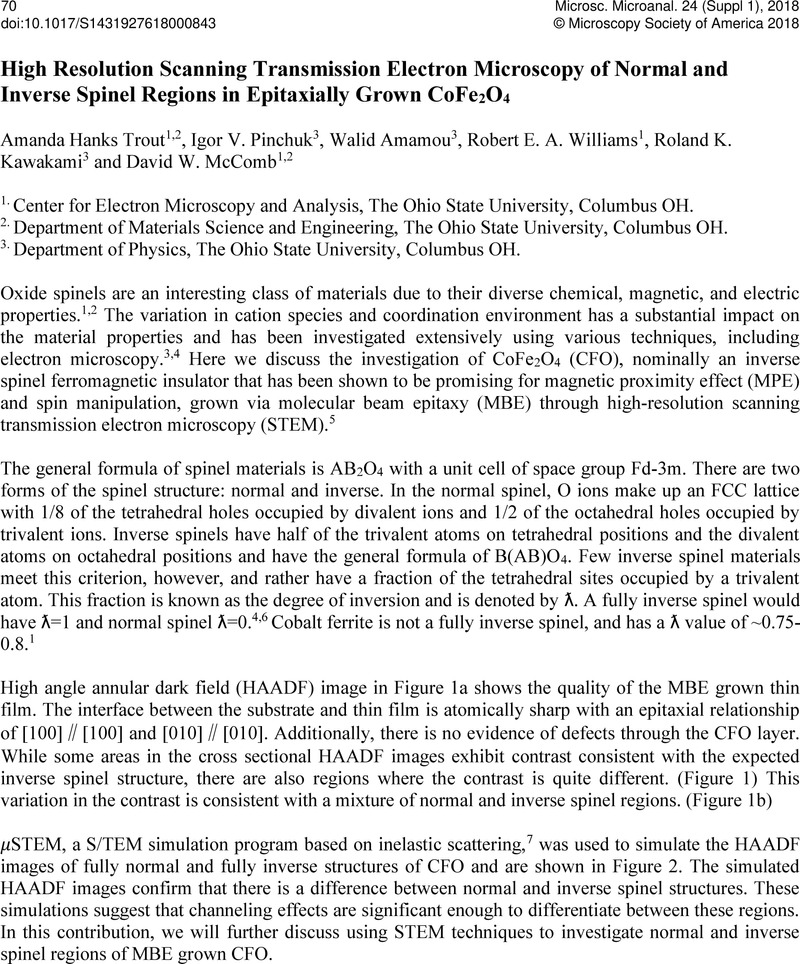
Image of the first page of this content. For PDF version, please use the ‘Save PDF’ preceeding this image.'
Type
Abstract
Copyright
© Microscopy Society of America 2018 

References

[1] Ferreira, T. A. S., et al, Solid State Sciences 5 2003) p. 383.Google Scholar
[2] Tatarchuk, T. R., et al, Journal of Alloys and Compounds 694 2017) p. 777.Google Scholar
[3] Docherty, F. T., et al, Ultramicroscopy 86 2001) p. 273.Google Scholar
[4] Tileli, V., et al, Nanoscale 7 2015) p. 218.Google Scholar
[5] Amamou, W., et al, Physical Review Materials 2 2018.Google Scholar
[6] Gobbi, G. C., et al, Chemistry Letters 14 1985) p. 771.Google Scholar
[7] Allen, L. J., et al, Ultramicroscopy 151 2015) p. 11.Google Scholar