Hostname: page-component-cd9895bd7-gbm5v Total loading time: 0 Render date: 2024-12-22T20:01:37.787Z Has data issue: false hasContentIssue false

Energy-Dispersive X-Ray Spectrum Simulation with NIST DTSA-II: Comparing Simulated and Measured Electron-Excited Spectra

Published online by Cambridge University Press:  02 September 2022
Dale E. Newbury*
Affiliation:
National Institute of Standards and Technology, Gaithersburg, MD 20899, USA
Nicholas W. M. Ritchie
Affiliation:
National Institute of Standards and Technology, Gaithersburg, MD 20899, USA
*
*Corresponding author: Dale E. Newbury, E-mail: [email protected]
Get access

Abstract

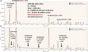
Electron-excited X-ray microanalysis with energy-dispersive spectrometry (EDS) proceeds through the application of the software that extracts characteristic X-ray intensities and performs corrections for the physics of electron and X-ray interactions with matter to achieve quantitative elemental analysis. NIST DTSA-II is an open-access, fully documented, and freely available comprehensive software platform for EDS quantification, measurement optimization, and spectrum simulation. Spectrum simulation with DTSA-II enables the prediction of the EDS spectrum from any target composition for a specified electron dose and for the solid angle and window parameters of the EDS spectrometer. Comparing the absolute intensities for measured and simulated spectra reveals correspondence within ±25% relative to K-shell and L-shell characteristic X-ray peaks in the range of 1–11 keV. The predicted M-shell intensity exceeds the measured value by a factor of 1.4–2.2 in the range 1–3 keV. The X-ray continuum (bremsstrahlung) generally agrees within ±10% over the range of 1–10 keV. Simulated EDS spectra are useful for developing an analytical strategy for challenging problems such as estimating trace detection levels.

Type
Software and Instrumentation
Copyright
Copyright © The Author(s), 2022. Published by Cambridge University Press on behalf of the Microscopy Society of America

Access options

Get access to the full version of this content by using one of the access options below. (Log in options will check for institutional or personal access. Content may require purchase if you do not have access.)

References

Goldstein, J, Newbury, D, Michael, J, Ritchie, N, Scott, J & Joy, D (2018). Scanning Electron Microscopy and X-Ray Microanalysis, 4th ed. New York: Springer.CrossRefGoogle Scholar
International Organization for Standardization (2003). standard ISO 22029:2003.Google Scholar
Newbury, D & Ritchie, N (2015 a). Review: Performing elemental microanalysis with high accuracy and high precision by scanning electron microscopy/silicon drift detector energy dispersive X-ray spectrometry (SEM/SDD-EDS). J Mats Sci 50, 493518.CrossRefGoogle Scholar
Newbury, DE & Ritchie, NWM (2015 b). Quantitative electron-excited X-Ray microanalysis of borides, carbides, nitrides, oxides, and fluorides with scanning electron microscopy/silicon drift detector energy-dispersive spectrometry (SEM/SDD-EDS) and DTSA-II. Microsc Microanal 21(2015), 13271340.CrossRefGoogle ScholarPubMed
Newbury, DE & Ritchie, NWM (2016 a). Electron-excited X-ray microanalysis at low beam energy: Almost always an adventure!. Micros Microanal 22, 735753.CrossRefGoogle ScholarPubMed
Newbury, DE & Ritchie, NWM (2016 b). Measurement of trace constituents by electron-excited X-ray microanalysis with energy-dispersive spectrometry. Microsc Microanal 22, 520535.CrossRefGoogle ScholarPubMed
Newbury, DE & Ritchie, NWM (2018). An iterative qualitative–quantitative sequential analysis strategy for electron-excited X-ray microanalysis with energy dispersive spectrometry: Finding the unexpected needles in the peak overlap haystack. Microsc Microanal 24, 350373.CrossRefGoogle ScholarPubMed
Pouchou, J-L & Pichoir, F (1991). Quantitative analysis of homogeneous or stratified microvolumes applying the model “PAP. In Electron Probe Quantitation, Heinrich, KFJ & Newbury, DE (Eds.), pp. 31. New York: Plenum.CrossRefGoogle Scholar
Ritchie, NW (2021). DTSA-II open access software for quantitative electron excited X-ray microanalysis with energy dispersive spectrometry. available for free, including tutorials, at the NIST. Available at https://www.nist.gov/services-resources/software/nist-dtsa-iiGoogle Scholar
Ritchie, NWM (2009). Spectrum simulation in DTSA-II. Microsc Microanal 15, 454468.CrossRefGoogle ScholarPubMed
Small, JA, Leigh, SD, Newbury, DE & Myklebust, RL (1987). Modeling of the bremsstrahlung radiation produced in pure-element targets by 10–40 keV electrons. J Appl Phys 61, 459469.CrossRefGoogle Scholar