Hostname: page-component-586b7cd67f-2brh9 Total loading time: 0 Render date: 2024-11-26T02:01:56.922Z Has data issue: false hasContentIssue false

Electron Microscopy as an Emerging Analytical Tool for Characterizing Biopharmaceuticals

Published online by Cambridge University Press:  23 September 2015

Bridget Carragher
Affiliation:
NanoImaging Services, Inc., San Diego, CAUSA
Anette Schneemann
Affiliation:
NanoImaging Services, Inc., San Diego, CAUSA
Joyce J. Sung
Affiliation:
NanoImaging Services, Inc., San Diego, CAUSA
Sean K. Mulligan
Affiliation:
NanoImaging Services, Inc., San Diego, CAUSA
Jeffrey A. Speir
Affiliation:
NanoImaging Services, Inc., San Diego, CAUSA
Kat On
Affiliation:
NanoImaging Services, Inc., San Diego, CAUSA
Joel Quispe
Affiliation:
NanoImaging Services, Inc., San Diego, CAUSA
Clinton S. Potter
Affiliation:
NanoImaging Services, Inc., San Diego, CAUSA

Abstract

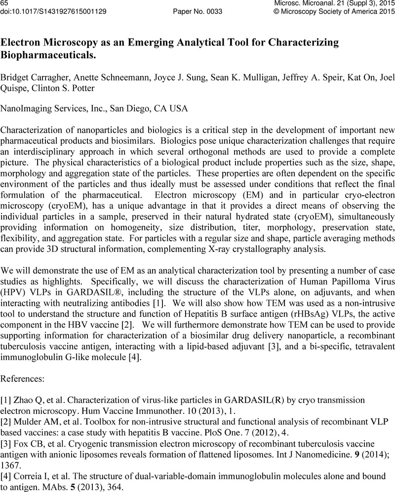
Image of the first page of this content. For PDF version, please use the ‘Save PDF’ preceeding this image.'
Type
Abstract
Copyright
Copyright © Microscopy Society of America 2015 

References

[1] Zhao, Q, et al. Characterization of virus-like particles in GARDASIL(R) by cryo transmission electron microscopy. Hum Vaccine Immunother 10 (2013), 1.Google Scholar
[2] Mulder, AM, et al. Toolbox for non-intrusive structural and functional analysis of recombinant VLP based vaccines: a case study with hepatitis B vaccine. PloS One 7 (2012), 4.CrossRefGoogle ScholarPubMed
[3] Fox, CB, et al. Cryogenic transmission electron microscopy of recombinant tuberculosis vaccine antigen with anionic liposomes reveals formation of flattened liposomes. Int J Nanomedicine 9 (2014), 1367.CrossRefGoogle ScholarPubMed
[4] Correia, I, et al. The structure of dual-variable-domain immunoglobulin molecules alone and bound to antigen. MAbs 5 (2013), 364.Google Scholar