Hostname: page-component-586b7cd67f-gb8f7 Total loading time: 0 Render date: 2024-11-23T01:05:33.939Z Has data issue: false hasContentIssue false

Differential Distribution of Flavonoids and Phenolic Acids in Leaves of Kalanchoe delagoensis Ecklon & Zeyher (Crassulaceae)

Published online by Cambridge University Press:  19 August 2020

Jamile Marques Casanova
Affiliation:
Plant Anatomy Laboratory, Department of Botany, Institute of Biology, Federal University of Rio de Janeiro, Rio de Janeiro21941-902, Brazil
Luana Beatriz dos Santos Nascimento
Affiliation:
Plant Anatomy Laboratory, Department of Botany, Institute of Biology, Federal University of Rio de Janeiro, Rio de Janeiro21941-902, Brazil
Livia Marques Casanova
Affiliation:
Laboratory of Chemistry for Bioactive Natural Products, Institute of Research on Natural Products, Federal University of Rio de Janeiro, Rio de Janeiro21941-902, Brazil
Marcos Vinicius Leal-Costa
Affiliation:
Federal Institute of Education, Science and Technology, Cabo Frio28909-971, Brazil
Sônia Soares Costa*
Affiliation:
Laboratory of Chemistry for Bioactive Natural Products, Institute of Research on Natural Products, Federal University of Rio de Janeiro, Rio de Janeiro21941-902, Brazil
Eliana Schwartz Tavares*
Affiliation:
Plant Anatomy Laboratory, Department of Botany, Institute of Biology, Federal University of Rio de Janeiro, Rio de Janeiro21941-902, Brazil
*
*Authors for correspondence: Sônia Soares Costa, E-mail: [email protected]; Eliana Schwartz Tavares, E-mail: [email protected]
Get access

Abstract

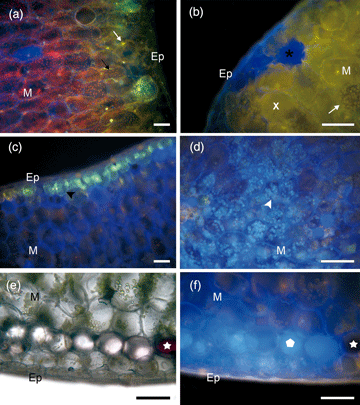
Kalanchoe delagoensis is adapted to intense solar irradiation, drought, and heat, partially due to the presence of phenols, important photo-protective compounds and antioxidants. This study aimed to evaluate the distribution of flavonoids and phenolic acid derivatives throughout the erect-tubular leaves of K. delagoensis. Specimens grown under sunny conditions were used for histochemical and high-performance liquid chromatography coupled with diode array detection (liquid HPLC-DAD) analysis. The NP (2-aminoethyl diphenylborinate) test suggested the presence of phenolic acids throughout the leaf blade below the epidermis and in chloroplasts, mainly in the leaf base. Flavonoids were detected specifically in chloroplasts, on the adaxial side of the middle third and at the leaf apex, near the meristematic cells. There was a tendency of flavonoid accumulation from the middle third to the apex, especially surrounding the gem, while phenolic acids were observed mainly in the base. This can be explained by the more exposed leaf apex and to the presence of apical buds (high production and regulation sites of ROS). The HPLC-DAD analysis showed different classes of flavonoids and phenolic acid derivatives in the leaf extracts, agreeing with the NP test results. This is the first time that the substitution of phenolic acids by flavonoids from the leaf base to the apex has been described.

Type
Micrographia
Copyright
Copyright © Microscopy Society of America 2020

Access options

Get access to the full version of this content by using one of the access options below. (Log in options will check for institutional or personal access. Content may require purchase if you do not have access.)

References

Abdel-Raouf, HS (2012). Anatomical traits of some species of Kalanchoe (Crassulaceae) and their taxonomic value. Ann Agric Sci 57(1), 7379.CrossRefGoogle Scholar
Agati, G, Azzarello, E, Pollastri, S & Tattini, M (2012). Flavonoids as antioxidants in plants: Location and functional significance. Plant Sci 196, 6776.CrossRefGoogle ScholarPubMed
Agati, G, Galardi, C, Gravano, E, Romani, A & Tattini, M (2002). Flavonoid distribution in tissues of Phillyrea latifolia L. leaves as estimated by microspectrofluorometry and multispectral fluorescence microimaging. Photochem Photobiol 76(3), 350360.2.0.CO;2>CrossRefGoogle ScholarPubMed
Alemanno, L, Ramos, T, Gargadenec, A, Andary, C & Ferriere, N (2003). Localization and identification of phenolic compounds in Theobroma cacao L. somatic embryogenesis. Ann Bot 92(4), 613–23.CrossRefGoogle ScholarPubMed
Asiedu-Gyekye, IJ, Antwi, DA, Bugyei, KA & Awortwe, C (2012). Comparative study of two Kalanchoe species: Total flavonoid and phenolic contents and antioxidant properties. African J Pure Appl Chem 6(5), 6573.Google Scholar
Balsamo, RA & Uribe, EG (1988). Leaf anatomy and ultrastructure of the Crassulacean-acid-metabolism plant Kalanchoe daigremontiana. Planta 173(2), 183189.CrossRefGoogle ScholarPubMed
Beveridge, C, Mathesius, U, Rose, R & Gresshoff, P (2007). Common regulatory themes in meristem development and whole-plant homeostasis. Curr Opin Plant Biol 10(1), 4451.10.1016/j.pbi.2006.11.011CrossRefGoogle ScholarPubMed
Bidel, LPR, Chomicki, G, Bonini, F, Mondolot, L, Soulé, J, Coumans, M, La Fisca, P, Baissac, Y, Petit, V, Loiseau, A, Cerovic, ZG, Gould, KS & Jay-Allemand, C (2015). Dynamics of flavonol accumulation in leaf tissues under different UV-B regimes in Centella asiatica (Apiaceae). Planta 242(3), 545559.CrossRefGoogle Scholar
Bogucka-Kocka, A, Zidorn, C, Kasprzycka, M, Szymczak, G & Szewczyk, K (2018). Phenolic acid content, antioxidant and cytotoxic activities of four Kalanchoë species. Saudi J Biol Sci 25(4), 622630.CrossRefGoogle ScholarPubMed
Boiteau, P & Allorge-Boiteau, L (1995). Kalanchoe (Crassulaceae) de Madagascar, Systématique, écophysiology et phytochimie. Paris: Karthala.Google Scholar
Brunetti, C, Di Ferdinando, M, Fini, A, Pollastri, S & Tattini, M (2013). Flavonoids as antioxidants and developmental regulators: Relative significance in plants and humans. Int J Mol Sci 14(2), 35403555.CrossRefGoogle ScholarPubMed
Bukatsch, F (1972). Bermerkungen zur doppelfarbung Astrablau-Safranin. Mikrokosmos 61, 255.Google Scholar
Casanova, LM, Da Silva, D, Sola-Penna, M, Camargo, LMM, Celestrini, DM, Tinoco, LW & Costa, SS (2014). Identification of chicoric acid as a hypoglycemic agent from Ocimum gratissimum leaf extract in a biomonitoring in vivo study. Fitoterapia 93, 132141.CrossRefGoogle Scholar
Castricini, SD (2004). Estudo fitoquímico de Kalanchoe fedtschenkoi (Crassulaceae) e avaliação de efeito de suas frações flavonoídicas em células tumorais. Master Thesis. Universidade Federal do Rio de Janeiro, Rio de Janeiro.Google Scholar
Chalker-Scott, L (1999). Environmental significance of anthocyanins in plant stress responses. Photochem Photobiol 70(1), 19.CrossRefGoogle Scholar
Chen, CH, Xu, H, Liu, XH, Zou, LS, Wang, M, Liu, ZX, Fu, XS, Zhao, H & Yan, Y (2017). Site-specific accumulation and dynamic change of flavonoids in Apocyni veneti Folium. Microsc Res Tech 80(12), 13151322.CrossRefGoogle ScholarPubMed
Chernetskyy, M & Weryszko-Chmielewska, E (2012). Structure of Kalanchoë pumila Bak. leaves (Crassulaceae DC). Acta Agrobot 61(2), 1124.CrossRefGoogle Scholar
Costa, SS, Muzitano, MF, Camargo, LMM & Coutinho, MAS (2008). Therapeutic potential of Kalanchoe species: Flavonoids and other secondary metabolites. Nat Prod Commun 3(12), 21512164.Google Scholar
Cruz, BP, Chedier, LM, Peixoto, PHP, Fabri, RL & Pimenta, DS (2012). Effects of light intensity on the distribution of anthocyanins in Kalanchoe brasiliensis Camb. and Kalanchoe pinnata (Lamk.) Pers. An Acad Bras Cienc 84(1), 211218.CrossRefGoogle ScholarPubMed
Donaldson, LA & Radotic, K (2013). Fluorescence lifetime imaging of lignin autofluorescence in normal and compression wood. J Microsc 251(2), 178187.CrossRefGoogle ScholarPubMed
Evert, RF (2013). Anatomia das Plantas de Esau: Meristemas, células e tecidos do corpo da planta – sua estrutura, função e desenvolvimento. São Paulo: Blucher.Google Scholar
Figueiredo, ACS, Barroso, JMG, Pedro, LMG & Ascensão, L (2007). Histoquímica e Citoquímica em Plantas: Princípios e Protocolos. Lisboa: Faculdade de Ciências - Universidade de Lisboa.Google Scholar
Gavin, NM & Durako, MJ (2012). Localization and antioxidant capacity of flavonoids in Halophila johnsonii in response to experimental light and salinity variation. J Exp Mar Bio Ecol 416–417, 3240.CrossRefGoogle Scholar
Gibson, AC (1996). Structure-Function Relations of Warm Desert Plants, 1st ed. Berlin: Springer.CrossRefGoogle Scholar
Gould, KS, Jay-Allemand, C, Logan, BA, Baissac, Y & Bidel, LPR (2018). When are foliar anthocyanins useful to plants? Re-evaluation of the photoprotection hypothesis using Arabidopsis thaliana mutants that differ in anthocyanin accumulation. Environ Exp Bot 154, 1122.CrossRefGoogle Scholar
Grünz, G, Haas, K, Soukup, S, Klingenspor, M, Kulling, SE, Daniel, H & Spanier, B (2012). Structural features and bioavailability of four flavonoids and their implications for lifespan-extending and antioxidant actions in C. elegans. Mech Ageing Dev 133(1), 110.CrossRefGoogle ScholarPubMed
Hatier, JHB & Gould, KS (2008). Anthocyanin function in vegetative organs. In Anthocyanins, Winefield, C, Davies, K & Gould, K (Eds.), pp. 119. New York, NY: Springer.Google ScholarPubMed
Holopainen, JK, Kivimäenpää, M & Julkunen-Tiitto, R (2018). New light for phytochemicals. Trends Biotechnol 36(1), 710.10.1016/j.tibtech.2017.08.009CrossRefGoogle ScholarPubMed
Huang, HC, Huang, GJ, Liaw, CC, Yang, CS, Yang, CP, Kuo, CL, Tseng, YH, Wang, SY, Chang, WT & Kuo, YH (2013). A new megastigmane from Kalanchoe tubiflora (Harvey) Hamet. Phytochem Lett 6(3), 379382.CrossRefGoogle Scholar
Johansen, DA (1940). Plant Microtechnique. New York, NY: Mcgraw-Hill Book Company.Google Scholar
Julkunen-Tiitto, R, Nenadis, N, Neugart, S, Robson, M, Agati, G, Vepsäläinen, J, Zipoli, G, Nybakken, L, Winkler, B & Jansen, MAK (2015). Assessing the response of plant flavonoids to UV radiation: An overview of appropriate techniques. Phytochem Rev 14(2), 273297.CrossRefGoogle Scholar
Korkina, LG (2007). Phenylpropanoids as naturally occurring antioxidants: From plant defense to human health. Cell Mol Biol 53(1), 1525.Google ScholarPubMed
Lang, M, Stober, F & Lichtenthaler, HK (1991). Fluorescence emission spectra of plant leaves and plant constituents. Radiat Environ Biophys 30, 333347.CrossRefGoogle ScholarPubMed
Livanos, P, Apostolakos, P & Galatis, B (2012). Plant cell division. Plant Signal Behav 7(7), 771778.10.4161/psb.20530CrossRefGoogle ScholarPubMed
Lopes-da-Silva, F, de Pascual-Teresa, S, Rivas-Gonzalo, J & Santos-Buelga, C (2002). Identification of anthocyanin pigments in strawberry (cv Camarosa) by LC using DAD and ESI-MS detection. Eur Food Res Technol 214(3), 248253.CrossRefGoogle Scholar
Lorenzi, H & Souza, VC (2012). Botânica sistemática: guia ilustrado para a identificação das famílias de fanerógamas nativas e exóticas no Brasil, baseado em APG III, 3rd ed. Nova Odessa: Instituto Plantarum.Google Scholar
Mabry, TJ, Markham, KR & Thomas, MB (1970). The Systematic Identification of Flavonoids, 1st ed. Berlin: Springer.CrossRefGoogle Scholar
Martins, GBC, Sucupira, RR & Suarez, PAZ (2015). A Química e as Cores. Rev Virtual Quim 7, 15081534.CrossRefGoogle Scholar
Matsuura, HN, de Costa, F, Yendo, ACA & Fett-Neto, AG (2013). Photoelicitation of bioactive secondary metabolites by ultraviolet radiation: Mechanisms, strategies, and applications. In Biotechnology for Medicinal Plants, Chandra, S, Lata, H & Varma, A (Eds.), pp. 171190. Berlin: Springer.CrossRefGoogle Scholar
McLusky, SR, Bennett, MH, Beale, MH, Lewis, MJ, Gaskin, P & Mansfield, JW (1999). Cell wall alterations and localized accumulation of feruloyl-3′-methoxytyramine in onion epidermis at sites of attempted penetration by Botrytis allii are associated with actin polarisation, peroxidase activity and suppression of flavonoid biosynthesis. Plant J 17(5), 523534.CrossRefGoogle Scholar
Melo, GO, Malvar, DC, Vanderlinde, FA, Rocha, FF, Pires, PA, Costa, EA, Matos, LG, Kaiser, CR & Costa, SS (2009). Antinociceptive and anti-inflammatory kaempferol glycosides from Sedum dendroideum. J Ethnopharmacol 124(2), 228232.CrossRefGoogle ScholarPubMed
Metcalfe, CR & Chalk, L (1950). Anatomy of the Dicotyledons, 1st ed. Oxford: Caredon Press.Google Scholar
Metcalfe, CR & Chalk, L (1979). Anatomy of the Dicotyledons, 2nd ed. Oxford: Caredon Press.Google Scholar
Mohan, SC, Balamurugan, V, Salini, ST & Rekha, R (2012). Metal ion chelating activity and hydrogen peroxide scavenging activity of medicinal plant Kalanchoe pinnata. J Chem Pharm Res 4(1), 197202.Google Scholar
Moreira, NS, Nascimento, LBS, Leal-Costa, MV & Tavares, ES (2012). Comparative anatomy of leaves of Kalanchoe pinnata and K. crenata in sun and shade conditions, as a support for their identification. Rev Bras Farmacogn 22(5), 929936.CrossRefGoogle Scholar
Muzitano, MF, Tinoco, LW, Guette, C, Kaiser, CR, Rossi-Bergmann, B & Costa, SS (2006). The antileishmanial activity assessment of unusual flavonoids from Kalanchoe pinnata. Phytochemistry 67(18), 20712077.CrossRefGoogle ScholarPubMed
Nelson, EA, Sage, TL & Sage, RF (2005). Functional leaf anatomy of plants with crassulacean acid metabolism. Funct Plant Biol 32(5), 409419.CrossRefGoogle ScholarPubMed
Pavia, DL, Lampman, GM & Kriz, GS (2010). Introdução à Espectroscopia, 4th ed. São Paulo: Cenage.Google Scholar
Quideau, S, Deffieux, D, Douat-Casassus, C & Pouységu, L (2011). Plant polyphenols: Chemical properties, biological activities, and synthesis. Angew Chemie Int 50(3), 586621.CrossRefGoogle ScholarPubMed
Reboredo, F & Lidon, FJC (2012). UV-B radiation effects on terrestrial plants – a perspective. Emirates J Food Agric 24(6), 502509.CrossRefGoogle Scholar
Robbins, RJ (2003). Phenolic acids in foods: An overview of analytical methodology. J Agric Food Chem 51(10), 28662887.CrossRefGoogle ScholarPubMed
Schnitzler, JP, Jungblut, TP, Werner, H, Kofferlein, M, Hutzler, UH, Schmelzer, E, Ernst, D, Langebartels, C & Sandermann, JRH (1996). Tissue localization of UV-B-screening pigments and of chalcone synthase mRNA in needles of Scots pine seedlings. New Phytol 132(2), 247258.CrossRefGoogle Scholar
Talamond, P, Verdeil, J-L & Conéjéro, G (2015). Secondary metabolite localization by autofluorescence in living plant cells. Molecules 20(3), 50245037.CrossRefGoogle ScholarPubMed
Tattini, M, Galardi, C, Pinelli, P, Massai, R, Remorini, D & Agati, G (2004). Differential accumulation of flavonoids and hydroxycinnamates in leaves of Ligustrum vulgare under excess light and drought stress. New Phytol 163(3), 547561.CrossRefGoogle ScholarPubMed
Tattini, M, Gravano, E, Pinelli, P, Mulinacci, N & Romani, A (2000). Flavonoids accumulate in leaves and glandular trichomes of Phillyrea latifolia exposed to excess solar radiation. New Phytol 148(1), 6977.CrossRefGoogle ScholarPubMed
Wagner, H & Bladt, S (2001). Plant Drug Analysis: A Thin Layer Chromatography Atlas, 2nd ed. München: Springer.Google Scholar
Wrolstad, RE, Acree, TE, Decker, EA, Penner, MH, Reid, DS, Schwartz, SJ, Shoemaker, CF, Smith, D & Sporns, P (2004). Handbook of Food Analytical Chemistry. Hoboken, NJ: John Wiley & Sons, Inc.CrossRefGoogle Scholar
Wrolstad, RE, Durst, RW & Lee, J (2005). Tracking color and pigment changes in anthocyanin products. Trends Food Sci Technol 16(9), 423428.CrossRefGoogle Scholar