Hostname: page-component-586b7cd67f-gb8f7 Total loading time: 0 Render date: 2024-11-22T19:45:19.527Z Has data issue: false hasContentIssue false

Development of an Energy-Sensitive Detector for the Atom Probe Tomography

Published online by Cambridge University Press:  20 September 2021
Christian Bacchi*
Affiliation:
Normandie Université, UNIROUEN, INSA Rouen, CNRS, Groupe de Physique des Matériaux, 76000 Rouen, France
Gérald Da Costa
Affiliation:
Normandie Université, UNIROUEN, INSA Rouen, CNRS, Groupe de Physique des Matériaux, 76000 Rouen, France
Emmanuel Cadel
Affiliation:
Normandie Université, UNIROUEN, INSA Rouen, CNRS, Groupe de Physique des Matériaux, 76000 Rouen, France
Fabien Cuvilly
Affiliation:
Normandie Université, UNIROUEN, INSA Rouen, CNRS, Groupe de Physique des Matériaux, 76000 Rouen, France
Jonathan Houard
Affiliation:
Normandie Université, UNIROUEN, INSA Rouen, CNRS, Groupe de Physique des Matériaux, 76000 Rouen, France
Charly Vaudolon
Affiliation:
Normandie Université, UNIROUEN, INSA Rouen, CNRS, Groupe de Physique des Matériaux, 76000 Rouen, France
Antoine Normand
Affiliation:
Normandie Université, UNIROUEN, INSA Rouen, CNRS, Groupe de Physique des Matériaux, 76000 Rouen, France
François Vurpillot
Affiliation:
Normandie Université, UNIROUEN, INSA Rouen, CNRS, Groupe de Physique des Matériaux, 76000 Rouen, France
*
*Corresponding author: Christian Bacchi, E-mail: [email protected]
Get access

Abstract

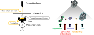
A position and energy-sensitive detector has been developed for atom probe tomography (APT) instruments in order to deal with some mass peak overlap issues encountered in APT experiments. Through this new type of detector, quantitative and qualitative improvements could be considered for critical materials with mass peak overlaps, such as nitrogen and silicon in TiSiN systems, or titanium and carbon in cemented carbide materials. This new detector is based on a thin carbon foil positioned on the front panel of a conventional MCP-DLD detector. According to several studies, it has been demonstrated that the impact of ions on thin carbon foils has the effect of generating a number of transmitted and reflected secondary electrons. The number generated mainly depends on both the kinetic energy and the mass of incident particles. Despite the fact that this phenomenon is well known and has been widely discussed for decades, no studies have been performed to date for using it as a means to discriminate particles energy. Therefore, this study introduces the first experiments on a potential new generation of APT detectors that would be able to resolve mass peak overlaps through the energy-sensitivity of thin carbon foils.

Type
Development and Computation
Copyright
Copyright © The Author(s), 2021. Published by Cambridge University Press on behalf of the Microscopy Society of America

Access options

Get access to the full version of this content by using one of the access options below. (Log in options will check for institutional or personal access. Content may require purchase if you do not have access.)

References

Allegrini, F, Ebert, RW & Funsten, HO (2016). Carbon foils for space plasma instrumentation. J Geophys Res: Space Phys 121, 39313950.CrossRefGoogle Scholar
Allegrini, F, Wimmer-Schweingruber, RF, Wurz, P & Bochsler, P (2003). Determination of low-energy ion-induced electron yields from thin carbon foils. Nucl Instrum Methods Phys Res B 211, 487494.CrossRefGoogle Scholar
Amirifar, N, Lardé, R, Talbot, E, Pareige, P, Rigutti, L, Mancini, L, Houard, J, Castro, C, Sallet, V, Zehani, E, Hassani, S, Sartel, C, Ziani, A & Portier, X (2015). Quantitative analysis of doped/undoped ZnO nanomaterials using laser assisted atom probe tomography: Influence of the analysis parameters. J Appl Phys 118, 215703.CrossRefGoogle Scholar
Bacchi, C (2020). New generation of position-sensitive detectors for the development of the atom probe tomography. Thesis. Available at https://tel.archives-ouvertes.fr/tel-03099387.Google Scholar
Bacchi, C, Da Costa, G & Vurpillot, F (2019). Spatial and compositional biases introduced by position sensitive detection systems in APT: A simulation approach. Microsc Microanal 25, 418424.CrossRefGoogle ScholarPubMed
Baragiola, RA (1993). Principles and mechanisms of ion induced electron emission. Nucl Instrum Methods Phys Res B 78, 223238.CrossRefGoogle Scholar
Barnstedt, J (2016). Advanced Practical Course Microchannel Plate Detectors. Tübingen, Germany: University of Tübingen.Google Scholar
Broderick, SR, Bryden, A, Suram, SK & Rajan, K (2013). Data mining for isotope discrimination in atom probe tomography. Ultramicroscopy 132, 121128.CrossRefGoogle ScholarPubMed
Buhr, H, Mendes, MB, Novotný, O, Schwalm, D, Berg, MH, Bing, D, Heber, O, Krantz, C, Orlov, DA, Rappaport, ML, Sorg, T, Stützel, J, Varju, J, Wolf, A & Zajfman, D (2010). Energy-sensitive imaging detector applied to the dissociative recombination of D2H+. Phys Rev A 81, 062702.CrossRefGoogle Scholar
Cerezo, A, Godfrey, TJ & Smith, GDW (1988). Application of a position-sensitive detector to atom probe microanalysis. Rev Sci Instrum 59, 862866.CrossRefGoogle Scholar
Chen, CY, Hun, CW, Chen, S-F, Chen, CC, Lin, JS, Johnson, SS, Noel, N, Juliely, N & Luo, Z (2015). Fabrication of nanoscale cesium iodide (CsI) scintillators for high-energy radiation detection. Rev Nanosci Nanotechnol 4, 2649.CrossRefGoogle Scholar
Chianelli, C, Ageron, P, Bouvet, JP, Karolak, M, Martin, S & Robert, JP (1988). Weakly ionizing charged particle detectors with high efficiency using transitory electronic secondary emission of porous CsI. Nucl Instrum Methods Phys Res A 273, 245256.CrossRefGoogle Scholar
Da Costa, G, Vurpillot, F, Bostel, A, Bouet, M & Deconihout, B (2005). Design of a delay-line position-sensitive detector with improved performance. Rev Sci Instrum 76, 013304.CrossRefGoogle Scholar
Da Costa, G, Wang, H, Duguay, S, Bostel, A, Blavette, D & Deconihout, B (2012). Advance in multi-hit detection and quantization in atom probe tomography. Rev Sci Instrum 83, 123709.CrossRefGoogle ScholarPubMed
Dawahre, N, Brewer, J, Shen, G, Harris, N, Wilbert, DS, Butler, L, Balci, S, Baughman, W, Kim, SM & Kung, P (2011). Nanoscale characteristics of single crystal zinc oxide nanowires. In 2011 11th IEEE International Conference on Nanotechnology, pp. 640–645. Portland, OR, USA: IEEE. Available at http://ieeexplore.ieee.org/document/6144626/ (accessed April 20, 2020).Google Scholar
Dennison, JR, Sim, A & Thomson, CD (2006). Evolution of the electron yield curves of insulators as a function of impinging electron fluence and energy. IEEE Trans Plasma Sci 34, 22042218.CrossRefGoogle Scholar
Drexler, CG & DuBois, RD (1996). Energy- and angle-differential yields of electron emission from thin carbon foils after fast proton impact. Phys Rev A 53, 16301637.CrossRefGoogle ScholarPubMed
Engberg, DLJ, Johnson, LJS, Jensen, J, Thuvander, M & Hultman, L (2018). Resolving mass spectral overlaps in atom probe tomography by isotopic substitutions – Case of TiSi15N. Ultramicroscopy 184, 5160.CrossRefGoogle ScholarPubMed
Fujii, G, Ukibe, M, Shiki, S & Ohkubo, M (2015). Development of array detectors with three-dimensional structure toward 1000 pixels of superconducting tunnel junctions. IEICE Trans Electron E98.C, 192195.CrossRefGoogle Scholar
Funsten, HO, Ritzau, SM, Harper, RW & Korde, R (2004). Fundamental limits to detection of low-energy ions using silicon solid-state detectors. Appl Phys Lett 84, 35523554.CrossRefGoogle Scholar
Gloeckler, G & Hsieh, KC (1979). Time-of-flight technique for particle identification at energies from 2–400 keV/nucleon. Nucl Instrum Methods 165, 537544.CrossRefGoogle Scholar
Goruganthu, RR & Wilson, WG (1984). Relative electron detection efficiency of microchannel plates from 0–3 keV. Rev Sci Instrum 55, 20302033.CrossRefGoogle Scholar
Hachenberg, O & Brauer, W (1959). Secondary electron emission from solids. In Advances in Electronics and Electron Physics, vol. 11, pp. 413499. Elsevier. Available at https://linkinghub.elsevier.com/retrieve/pii/S0065253908609993 (accessed May 29, 2021).Google Scholar
Hasselkamp, D (1992). Kinetic electron emission from solid surfaces under ion bombardment. In Particle Induced Electron Emission II, vol. 123, Springer Tracts in Modern Physics, Hasselkamp, D, Rothard, H, Groeneveld, K-O, Kemmler, J, Varga, P & Winter, H (Eds.), pp. 195. Berlin, Heidelberg: Springer Berlin Heidelberg. Available at http://link.springer.com/10.1007/BFb0038298 (accessed April 16, 2020).CrossRefGoogle Scholar
Hill, AG, Buechner, WW, Clark, JS & Fisk, JB (1939). The emission of secondary electrons under high energy positive ion bombardment. Phys Rev 55, 463470.CrossRefGoogle Scholar
Jackson, WJ (1927). Secondary emission from Mo due to bombardment by high speed positive ions of the alkali metals. Phys Rev 30, 473478.CrossRefGoogle Scholar
Jagutzki, O, Mergel, V, Ullmann-Pfleger, K, Spielberger, L, Spillmann, U, Dörner, R & Schmidt-Böcking, H (2002). A broad-application microchannel-plate detector system for advanced particle or photon detection tasks: Large area imaging, precise multi-hit timing information and high detection rate. Nucl Instrum Methods Phys Res A 477, 244249.CrossRefGoogle Scholar
Kelly, TF (2011). Kinetic-energy discrimination for atom probe tomography: Review article. Microsc Microanal 17, 114.CrossRefGoogle Scholar
Kirchhofer, R, Diercks, DR, Gorman, BP, Ihlefeld, JF, Kotula, PG, Shelton, CT & Brennecka, GL (2014). Quantifying compositional homogeneity in Pb(Zr,Ti)O3 using atom probe tomography Green, D. J. (Ed.). J Am Ceram Soc 97, 26772697.CrossRefGoogle Scholar
Klein, HJ (1965). Ausbeuten und winkelverteilungen der durch edelgasionen an reinen wolframoberflächen ausgelösten sekundärelektronen. Z Phys 188, 7892.CrossRefGoogle Scholar
Kuznetsov, AV, Veldhuizen, EJ, Westerberg, L, Lyapin, VG, Aleklett, K, Loveland, W, Bondorf, J, Jakobsson, B, Whitlow, HJ & El Bouanani, M (2000). A compact ultra-high vacuum (UHV) compatible instrument for time of flight–energy measurements of slow heavy reaction products. Nucl Instrum Methods Phys Res A 452, 525532.CrossRefGoogle Scholar
Ladislas Wiza, J (1979). Microchannel plate detectors. Nucl Instrum Methods 162, 587601.CrossRefGoogle Scholar
La Fontaine, A, Zavgorodniy, A, Liu, H, Zheng, R, Swain, M & Cairney, J (2016). Atomic-scale compositional mapping reveals Mg-rich amorphous calcium phosphate in human dental enamel. Sci Adv 2, e1601145.CrossRefGoogle ScholarPubMed
Maier-Komor, P (1993). Carbon foils for nuclear accelerator experiments. Nucl Instrum Methods Phys Res B 79, 841844.CrossRefGoogle Scholar
Marceau, RKW, Ceguerra, AV, Breen, AJ, Raabe, D & Ringer, SP (2015). Quantitative chemical-structure evaluation using atom probe tomography: Short-range order analysis of Fe–Al. Ultramicroscopy 157, 1220.CrossRefGoogle ScholarPubMed
Martin, AJ, Wei, Y & Scholze, A (2018). Analyzing the channel dopant profile in next-generation FinFETs via atom probe tomography. Ultramicroscopy 186, 104111.CrossRefGoogle ScholarPubMed
McCracken, GM (1975). The behaviour of surfaces under ion bombardment. Rep Prog Phys 38, 241327.CrossRefGoogle Scholar
Meisenkothen, F, Steel, EB, Prosa, TJ, Henry, KT & Prakash Kolli, R (2015). Effects of detector dead-time on quantitative analyses involving boron and multi-hit detection events in atom probe tomography. Ultramicroscopy 159, 101111.CrossRefGoogle ScholarPubMed
Miller, MK (1987). The effects of local magnification and trajectory aberrations on atom probe analysis. J Phys Colloques 48, C6-565C6-570.CrossRefGoogle Scholar
Miller, MK & Forbes, RG (2014). Atom-Probe Tomography. Boston, MA: Springer US. Available at http://link.springer.com/10.1007/978-1-4899-7430-3 (accessed July 9, 2018).CrossRefGoogle Scholar
Miodownik, M (2015). Materials for the 21st century: What will we dream up next? MRS Bull 40, 11881197.CrossRefGoogle Scholar
Montagnoli, G, Stefanini, AM, Trotta, M, Beghini, S, Bettini, M, Scarlassara, F, Schiavon, V, Corradi, L, Behera, BR, Fioretto, E, Gadea, A, Latina, A, Szilner, S, Donà, L, Rigato, M, Kondratiev, NA, Chizhov, AY, Kniajeva, G, Kozulin, EM, Pokrovskiy, IV, Voskressensky, VM & Ackermann, D (2005). The large-area micro-channel plate entrance detector of the heavy-ion magnetic spectrometer PRISMA. Nucl Instrum Methods Phys Res A 547, 455463.CrossRefGoogle Scholar
Müller, A, Djurić, N, Dunn, GH & Belić, DS (1986). Absolute detection efficiencies of microchannel plates for 0.1–2.3 keV electrons and 2.1–4.4 keV Mg+ ions. Rev Sci Instrum 57, 349353.CrossRefGoogle Scholar
Murdock, JW & Miller, GH (1995). Secondary electron emission due to positive ion bombardment. Available at https://lib.dr.iastate.edu/ameslab_iscreports/106.Google Scholar
Ohkubo, M, Shigetomo, S, Ukibe, M, Fujii, G & Matsubayashi, N (2014). Superconducting tunnel junction detectors for analytical sciences. IEEE Trans Appl Supercond 24, 1–8.CrossRefGoogle Scholar
Oliphant, MLE (1930). The liberation of electrons from metal surfaces by positive ions. Part I. Experimental. Proc R Soc A: Math Phys Eng Sci 127, 373387.Google Scholar
Palmberg, PW (1967). Secondary emission studies on Ge and Na-covered Ge. J Appl Phys 38, 21372147.CrossRefGoogle Scholar
Park, S, Jung, W & Park, C (2013). Distribution of nickel in zinc oxide nanowalls on sapphire substrate investigated using atom probe tomography. Scr Mater 68, 10001003.CrossRefGoogle Scholar
Peng, Z, Vurpillot, F, Choi, P-P, Li, Y, Raabe, D & Gault, B (2018). On the detection of multiple events in atom probe tomography. Ultramicroscopy 189, 5460.CrossRefGoogle ScholarPubMed
Rothard, H, Caraby, C, Cassimi, A, Gervais, B, Grandin, J-P, Jardin, P, Jung, M, Billebaud, A, Chevallier, M, Groeneveld, K-O & Maier, R (1995). Target-thickness-dependent electron emission from carbon foils bombarded with swift highly charged heavy ions. Phys Rev A 51, 30663078.CrossRefGoogle ScholarPubMed
Rothard, H, Jung, M, Caron, M, Grandin, J-P, Gervais, B, Billebaud, A, Clouvas, A & Wünsch, R (1998). Strong projectile-dependent forward-backward asymmetry of electron ejection by swift heavy ions in solids. Phys Rev A 57, 36603664.CrossRefGoogle Scholar
Rothard, H, Kroneberger, K, Clouvas, A, Veje, E, Lorenzen, P, Keller, N, Kemmler, J, Meckbach, W & Groeneveld, K-O (1990). Secondary-electron yields from thin foils: A possible probe for the electronic stopping power of heavy ions. Phys Rev A 41, 25212535.CrossRefGoogle ScholarPubMed
Rymzhanov, RA, Medvedev, NA & Volkov, AE (2015). Electron emission from silicon and germanium after swift heavy ion impact: Electron emission from Si and Ge after swift heavy ion impact. Phys Status Solidi B 252, 159164.CrossRefGoogle Scholar
Šaro, Š, Janik, R, Hofmann, S, Folger, H, Heßberger, FP, Ninov, V, Schött, HJ, Kabachenko, AP, Popeko, AG & Yeremin, AV (1996). Large size foil-microchannel plate timing detectors. Nucl Instrum Methods Phys Res A 381, 520526.CrossRefGoogle Scholar
Schou, J (1980). Transport theory for kinetic emission of secondary electrons from solids. Phys Rev B 22, 21412174.CrossRefGoogle Scholar
Seol, J-B, Raabe, D, Choi, P, Park, H-S, Kwak, J-H & Park, C-G (2013). Direct evidence for the formation of ordered carbides in a ferrite-based low-density Fe–Mn–Al–C alloy studied by transmission electron microscopy and atom probe tomography. Scr Mater 68, 348353.CrossRefGoogle Scholar
Shapira, D, Lewis, TA & Hulett, LD (2000). A fast and accurate position-sensitive timing detector based on secondary electron emission. Nucl Instrum Methods Phy Res A 454, 409420.CrossRefGoogle Scholar
Sternglass, EJ (1957). Theory of secondary electron emission by high-speed ions. Phys Rev 108, 112.CrossRefGoogle Scholar
Thuvander, M, Weidow, J, Angseryd, J, Falk, LKL, Liu, F, Sonestedt, M, Stiller, K & Andrén, H-O (2011). Quantitative atom probe analysis of carbides. Ultramicroscopy 111, 604608.CrossRefGoogle ScholarPubMed
Töglhofer, K, Aumayr, F & Winter, HP (1993). Ion-induced electron emission from metal surfaces — Insights from the emission statistics. Surf Sci 281, 143152.CrossRefGoogle Scholar
Tomita, S, Yoda, S, Uchiyama, R, Ishii, S, Sasa, K, Kaneko, T & Kudo, H (2006). Nonadditivity of convoy- and secondary-electron yields in the forward-electron emission from thin carbon foils under irradiation of fast carbon-cluster ions. Phys Rev A 73, 060901.CrossRefGoogle Scholar
Tsong, TT (1990). Atom-Probe Field Ion Microscopy: Field Ion Emission and Surfaces and Interfaces at Atomic Resolution. Cambridge: Cambridge University Press. Available at http://ebooks.cambridge.org/ref/id/CBO9780511599842 (accessed July 9, 2018).CrossRefGoogle Scholar
Vurpillot, F (2016). Field ion emission mechanisms. In Atom Probe Tomography. pp. 1772. Elsevier. Available at http://linkinghub.elsevier.com/retrieve/pii/B9780128046470000024 (accessed July 9, 2018).CrossRefGoogle Scholar
Vurpillot, F, Bostel, A & Blavette, D (2000). Trajectory overlaps and local magnification in three-dimensional atom probe. Appl Phys Lett 76, 31273129.CrossRefGoogle Scholar
Ward, BW, Shaver, DC & Ward, ML (1985). Repair of Photomasks with Focussed Ion Beams. Blais, PD (Ed.), p. 110. Santa Clara. Available at http://proceedings.spiedigitallibrary.org/proceeding.aspx?doi=10.1117/12.947491 (accessed January 30, 2020).Google Scholar
Weathers, LW & Tsang, MB (1996). Fabrication of thin scintillator foils. Nucl Instrum Methods Phys Res A 381, 567568.CrossRefGoogle Scholar
Wortmann, M, Ludwig, A, Meijer, J, Reuter, D & Wieck, AD (2013). High-resolution mass spectrometer for liquid metal ion sources. Rev Sci Instrum 84, 093305.CrossRefGoogle ScholarPubMed
Ziegler, JF & Biersack, JP (1985). The stopping and range of ions in matter. In Treatise on Heavy-Ion Science: Volume 6: Astrophysics, Chemistry, and Condensed Matter, Bromley, DA (Ed.), pp. 93129. Boston, MA: Springer US. Available at https://doi.org/10.1007/978-1-4615-8103-1_3 (accessed January 30, 2020).Google Scholar
Ziegler, JF, Ziegler, MD & Biersack, JP (2010). SRIM – The stopping and range of ions in matter (2010). Nucl Instrum Methods Phys Res B 268, 18181823.CrossRefGoogle Scholar