Hostname: page-component-586b7cd67f-tf8b9 Total loading time: 0 Render date: 2024-11-22T19:50:46.389Z Has data issue: false hasContentIssue false

Correlative Approach for Atom Probe Sample Preparation of Interfaces Using Plasma Focused Ion Beam Without Lift-Out

Published online by Cambridge University Press:  20 April 2021

Vitor Vieira Rielli
Affiliation:
School of Materials Science & Engineering, UNSW, Sydney, NSW 2052, Australia
Felix Theska
Affiliation:
School of Materials Science & Engineering, UNSW, Sydney, NSW 2052, Australia
Sophie Primig*
Affiliation:
School of Materials Science & Engineering, UNSW, Sydney, NSW 2052, Australia
*
*Author for correspondence: Sophie Primig, E-mail: [email protected]
Get access

Abstract

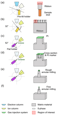
Plasma focused ion beam microscopy (PFIB) is a recent nanofabrication technique that is suitable for site-specific atom probe sample preparation. Higher milling rates and fewer artifacts make it superior to Ga+ FIBs for the preparation of samples where large volumes of material must be removed, for example, when trying to avoid lift-out techniques. Transmission Kikuchi diffraction (TKD) is a method that has facilitated phase identification and crystallographic measurements in such electron transparent samples. We propose a procedure for preparing atom probe tomography (APT) tips from mechanically prepared ribbons by using PFIB. This is highly suitable for the preparation of atom probe tips of interfaces such as interphase boundaries from challenging materials where lift-out tips easily fracture. Our method, in combination with TKD, allows the positioning of regions of interest such as interfaces close to the apex of the tip. We showcase the efficacy of the proposed method in a case study on Alloy 718, where the interface between γ-matrix and δ-phase has not been yet extensively explored through APT due to preparation challenges. Results show depletion of γ″-precipitates near the γ/δ interface. A quantitative evaluation of the composition of phases in the bulk versus near the interface is achieved.

Type
Development and Computation
Copyright
Copyright © The Author(s), 2021. Published by Cambridge University Press on behalf of the Microscopy Society of America

Access options

Get access to the full version of this content by using one of the access options below. (Log in options will check for institutional or personal access. Content may require purchase if you do not have access.)

References

Altmann, F & Young, RJ (2014). Site-specific metrology, inspection, and failure analysis of three-dimensional interconnects using focused ion beam technology. J Micro/Nanolith MEMS, MOEMS 13, 011202.CrossRefGoogle Scholar
Azadian, S, Wei, LY & Warren, R (2004). Delta phase precipitation in inconel 718. Mater Charact 53, 716.CrossRefGoogle Scholar
Babinsky, K, De Kloe, R, Clemens, H & Primig, S (2014). A novel approach for site-specific atom probe specimen preparation by focused ion beam and transmission electron backscatter diffraction. Ultramicroscopy 144, 918.CrossRefGoogle ScholarPubMed
Baillet, J, Gavarini, S, Millard-Pinard, N, Garnier, V, Peaucelle, C, Jaurand, X, Duranti, A, Bernard, C, Rapegno, R & Cardinal, S (2018). Surface damage on polycrystalline β-SiC by xenon ion irradiation at high fluence. J Nucl Mater 503, 140150.CrossRefGoogle Scholar
Blum, I, Cuvilly, F & Lefebvre-Ulrikson, W (2016). Atom probe sample preparation. In Atom Probe Tomography, Lefebvre, W, Vurpillot, F & Sauvage, X (Eds.), pp. 97121. Oxford, UK: Academic Press.CrossRefGoogle Scholar
Breen, AJ, Babinsky, K, Day, AC, Eder, K, Oakman, CJ, Trimby, PW, Primig, S, Cairney, JM & Ringer, SP (2017). Correlating atom probe crystallographic measurements with transmission Kikuchi diffraction data. Microsc Microanal 23, 279290.CrossRefGoogle ScholarPubMed
Burke, MG & Miller, MK (1990). Grain boundary intermetallic phases in Alloy 718. MRS Proc 186, 215218.CrossRefGoogle Scholar
Burke, MG & Miller, MK (1991). Precipitation in Alloy 718: A combined Al3M and apfim investigation. Superalloys 718, 337350.CrossRefGoogle Scholar
Burnett, TL, Kelley, R, Winiarski, B, Contreras, L, Daly, M, Gholinia, A, Burke, MG & Withers, PJ (2016). Large volume serial section tomography by Xe plasma FIB dual beam microscopy. Ultramicroscopy 161, 119129. doi:10.1016/j.ultramic.2015.11.001.CrossRefGoogle ScholarPubMed
Chen, YS, Lu, H, Liang, J, Rosenthal, A, Liu, H, Sneddon, G, McCarroll, I, Zhao, Z, Li, W, Guo, A & Cairney, JM (2020). Observation of hydrogen trapping at dislocations, grain boundaries, and precipitates. Science 367, 171175.CrossRefGoogle ScholarPubMed
Cozar, R & Pineau, A (1973). Morphology of y′ and y″ precipitates and thermal stability of inconel 718 type alloys. Metall Trans 4, 4759.CrossRefGoogle Scholar
Eder, K, Bhatia, V, Van Leer, B & Cairney, JM (2019). Using a plasma FIB equipped with Xe, N2, O2 and Ar for atom probe sample preparation–ion implantation and success rates. Microsc Microanal 25, 316317.CrossRefGoogle Scholar
Estivill, R, Audoit, G, Barnes, J-P, Grenier, A & Blavette, D (2016). Preparation and analysis of atom probe tips by xenon focused ion beam milling. Microsc Microanal 22, 576.CrossRefGoogle ScholarPubMed
Gault, B, Moody, MP, Cairney, JM & Ringer, SP (2012). Atom Probe Microscopy. New York: Springer Science & Business Media.CrossRefGoogle Scholar
Halpin, JE, Webster, RWH, Gardner, H, Moody, MP, Bagot, PAJ & Maclaren, DA (2019). An in-situ approach for preparing atom probe tomography specimens by xenon plasma-focussed ion beam. Ultramicroscopy 202, 121127. doi:10.1016/j.ultramic.2019.04.005.CrossRefGoogle ScholarPubMed
Holzer, L & Cantoni, M (2012. Review of FIB tomography. Nanofabrication Using Focused Ion and Electron beams. In Principles and Applications, Utke, I, Moshkalev, S & Russell, P (Eds.), pp. 410435.Oxford, UK. Oxford University.Google Scholar
Jinqiao, L, Ranming, N, Ji, G, Cabral, M, Song, M & Xiaozhou, L (2020). Effect of ion irradiation introduced by focused ion-beam milling on the mechanical behaviour of sub-micron-sized samples. Sci Rep 10.10324.Google Scholar
Keller, RR & Geiss, RH (2012). Transmission EBSD from 10 nm domains in a scanning electron microscope. J Microsc 245, 245251.CrossRefGoogle Scholar
Kellogg, S, Schampers, R, Zhang, S, Graupera, A, Miller, T, Laur, WD & Dirriwachter, A (2010). High throughput sample preparation and analysis using an inductively coupled plasma (ICP) focused Ion beam source. Microsc Microanal 16, 222223.CrossRefGoogle Scholar
Kirman, I & Warrington, DH (1970). The precipitation of Ni3Nb phases in a Ni-Fe-Cr-Nb alloy. Metall Trans 1, 26672675.CrossRefGoogle Scholar
Krakauer, BW & Seidman, DN (1992). Systematic procedures for atom-probe field-ion microscopy studies of grain boundary segregation. Rev Sci Instrum 63, 40714079.CrossRefGoogle Scholar
Lacaze, J, Dehmas, M, Niang, A & Viguier, B (2011). TEM study of high-temperature precipitation of delta phase in inconel 718 alloy. Adv Mater Sci Eng 2011.19.Google Scholar
Lawrence, D, Thompson, K & Larson, DJ (2006). Site-specific specimen preparation technique for atom probe analysis of grain boundaries. Microsc Microanal 12, 17401741.CrossRefGoogle Scholar
Li, J, Malis, T & Dionne, S (2006). Recent advances in FIB-TEM specimen preparation techniques. Mater Charact 57, 6470.CrossRefGoogle Scholar
Liu, J, Lozano-Perez, S, Wilkinson, AJ & Grovenor, CRM (2019). On the depth resolution of transmission Kikuchi diffraction (TKD) analysis. Ultramicroscopy 205, 512.CrossRefGoogle ScholarPubMed
Marquis, EA, Geiser, BP, Prosa, TJ & Larson, DJ (2011). Evolution of tip shape during field evaporation of complex multilayer structures. J Microsc 241, 225233.CrossRefGoogle ScholarPubMed
McCarroll, IE, Bagot, PAJ, Devaraj, A, Perea, DE & Cairney, JM (2020). New frontiers in atom probe tomography: A review of research enabled by cryo and/or vacuum transfer systems. Mater Today Adv 7, 100090.CrossRefGoogle ScholarPubMed
Miller, MK (2001). Contributions of atom probe tomography to the understanding of nickel-based superalloys. Micron 32, 757764.CrossRefGoogle Scholar
Miller, MK, Horton, JA, Cao, WD & Kennedy, RL (1996). Characterization of the effects of boron and phosphorus additions to the nickel-based superalloy 718. Journal de Physique IV 6, 16.Google Scholar
Miller, MK, Russell, KF, Thompson, K, Alvis, R & Larson, DJ (2007). Review of atom probe FIB-based specimen preparation methods. Microsc Microanal 13, 428436.CrossRefGoogle ScholarPubMed
Miller, MK, Russell, KF, Thompson, K, Alvis, R & Larson, DJ (2019). Review of Atom Probe FIB-Based Specimen Preparation Methods. doi:10.1017/S1431927607070845.CrossRefGoogle Scholar
Nalawade, SA, Sundararaman, M, Singh, JB, Verma, A & Kishore, R (2010). Precipitation of γ′ phase in δ-precipitated Alloy 718 during deformation at elevated temperatures. Mater Sci Eng A 527, 29062909. doi:10.1016/j.msea.2010.01.006.CrossRefGoogle Scholar
Ping, DH, Gu, YF, Cui, CY & Harada, H (2007). Grain boundary segregation in a Ni-Fe-based (Alloy 718) superalloy. Mater Sci Eng A 456, 99102.CrossRefGoogle Scholar
Quist, WE, Taggart, R & Polonis, DH (1971). The influence of iron and aluminum on the precipitation of metastable Ni3Nb phases in the Ni-Nb system. Metall Trans 2, 825832.CrossRefGoogle Scholar
Reed, RC (2006). The Superalloys: Fundamentals and Applications. Cambridge: Cambridge University Press.CrossRefGoogle Scholar
Saxey, DW, Cairney, JM, McGrouther, D, Honma, T & Ringer, SP (2007). Atom probe specimen fabrication methods using a dual FIB/SEM. Ultramicroscopy 107, 756760.CrossRefGoogle ScholarPubMed
Sneddon, GC, Trimby, PW & Cairney, JM (2016). Transmission Kikuchi diffraction in a scanning electron microscope: A review. Mater Sci Eng R: Rep 110, 112. doi:10.1016/j.mser.2016.10.001.CrossRefGoogle Scholar
Sun, WR, Guo, SR, Lee, JH, Park, NK, Yoo, YS, Choe, SJ & Hu, ZQ (1998). Effects of phosphorus on the δ-Ni3Nb phase precipitation and the stress rupture properties in Alloy 718. Mater Sci Eng A 247, 173179.CrossRefGoogle Scholar
Sundararaman, M, Mukhopadhyay, P & Banerjee, S (1988). Precipitation of the δ-Ni3Nb phase in two nickel base superalloys. Metall Trans A 19, 453465.CrossRefGoogle Scholar
Tarzimoghadam, Z, Rohwerder, M, Merzlikin, SV, Bashir, A, Yedra, L, Eswara, S, Ponge, D & Raabe, D (2016). Multi-scale and spatially resolved hydrogen mapping in a Ni–Nb model alloy reveals the role of the δ phase in hydrogen embrittlement of Alloy 718. Acta Mater 109, 6981.CrossRefGoogle Scholar
Theska, F, Ringer, SP & Primig, S (2019). Atom probe microscopy of strengthening effects in Alloy 718. Microsc Microanal 25.470480.CrossRefGoogle ScholarPubMed
Theska, F, Stanojevic, A, Oberwinkler, B & Primig, S (2020). Microstructure-property relationships in directly aged Alloy 718 turbine disks. Mater Sci Eng A 776, 138967. doi:10.1016/j.msea.2020.138967.CrossRefGoogle Scholar
Theska, F, Stanojevic, A, Oberwinkler, B, Ringer, SP & Primig, S (2018). On conventional versus direct ageing of Alloy 718. Acta Mater 156, 116124.CrossRefGoogle Scholar
Thompson, K, Lawrence, D, Larson, DJ, Olson, JD, Kelly, TF & Gorman, B (2007). In situ site-specific specimen preparation for atom probe tomography. Ultramicroscopy 107, 131139.CrossRefGoogle ScholarPubMed
Trimby, PW (2012). Orientation mapping of nanostructured materials using transmission Kikuchi diffraction in the scanning electron microscope. Ultramicroscopy 120, 1624.CrossRefGoogle ScholarPubMed
Turnbull, A, Ballinger, RG, Hwang, IS, Morra, MM, Psaila-Dombrowski, M & Gates, RM (1992). Hydrogen transport in nickel-base alloys. Metall Trans A 23, 32313244.CrossRefGoogle Scholar
Vander Voort, GF (1999). Metallography, Principles and Practice. Materials Park, OH: ASM International.Google Scholar
Viskari, L & Stiller, K (2011). Atom probe tomography of Ni-base superalloys Allvac 718Plus and Alloy 718. Ultramicroscopy 111, 652658. doi:10.1016/j.ultramic.2011.01.015.CrossRefGoogle ScholarPubMed
Vurpillot, F, Larson, D & Cerezo, A (2004). Improvement of multilayer analyses with a three-dimensional atom probe. Surf Interface Anal 36, 552558.CrossRefGoogle Scholar
Wei, Y, Peng, Z, Kühbach, M, Breen, A, Legros, M, Larranaga, M, Mompiou, F & Gault, B (2019). 3D nanostructural characterisation of grain boundaries in atom probe data utilising machine learning methods. PLoS One 14, 119.CrossRefGoogle ScholarPubMed
White, N, Eder, K, Byrnes, J, Cairney, JM & McCarroll, IE (2021). Laser ablation sample preparation for atom probe tomography and transmission electron microscopy. Ultramicroscopy 220, 113161.CrossRefGoogle ScholarPubMed
Wright, SI, Nowell, MM, de Kloe, R, Camus, P & Rampton, T (2015). Electron imaging with an EBSD detector. Ultramicroscopy 148, 132145.CrossRefGoogle ScholarPubMed
Zhang, Z, Moore, KL, McMahon, G, Morana, R & Preuss, M (2019). On the role of precipitates in hydrogen trapping and hydrogen embrittlement of a nickel-based superalloy. Corros Sci 146, 5869.CrossRefGoogle Scholar
Zhong, X, Wade, CA, Withers, PJ, Zhou, X, Cai, C, Haigh, SJ & Burke, MG (2020). Comparing Xe+ pFIB and Ga+ FIB for TEM sample preparation of Al alloys: Minimising FIB-induced artefacts. J Microsc.112.Google ScholarPubMed
Ziegler, JF & Biersack, JP (1985). The stopping and range of ions. In Treatise on Heavy-Ion Science: Astrophysics, Chemistry, and Condensed Matter, Bromley, MDA (Ed.), vol. 6. pp. 93129. Boston, MA: Springer.CrossRefGoogle Scholar