Hostname: page-component-586b7cd67f-t7czq Total loading time: 0 Render date: 2024-11-27T00:28:55.351Z Has data issue: false hasContentIssue false

Choice of Specimen Thickness in Axial Bright-Field STEM Tomography of Cells

Published online by Cambridge University Press:  25 July 2016

Qianping He
Affiliation:
National Institute of Biomedical Imaging and Bioengineering, NIH, Bethesda, MD 20892
Richard D. Leapman
Affiliation:
National Institute of Biomedical Imaging and Bioengineering, NIH, Bethesda, MD 20892

Abstract

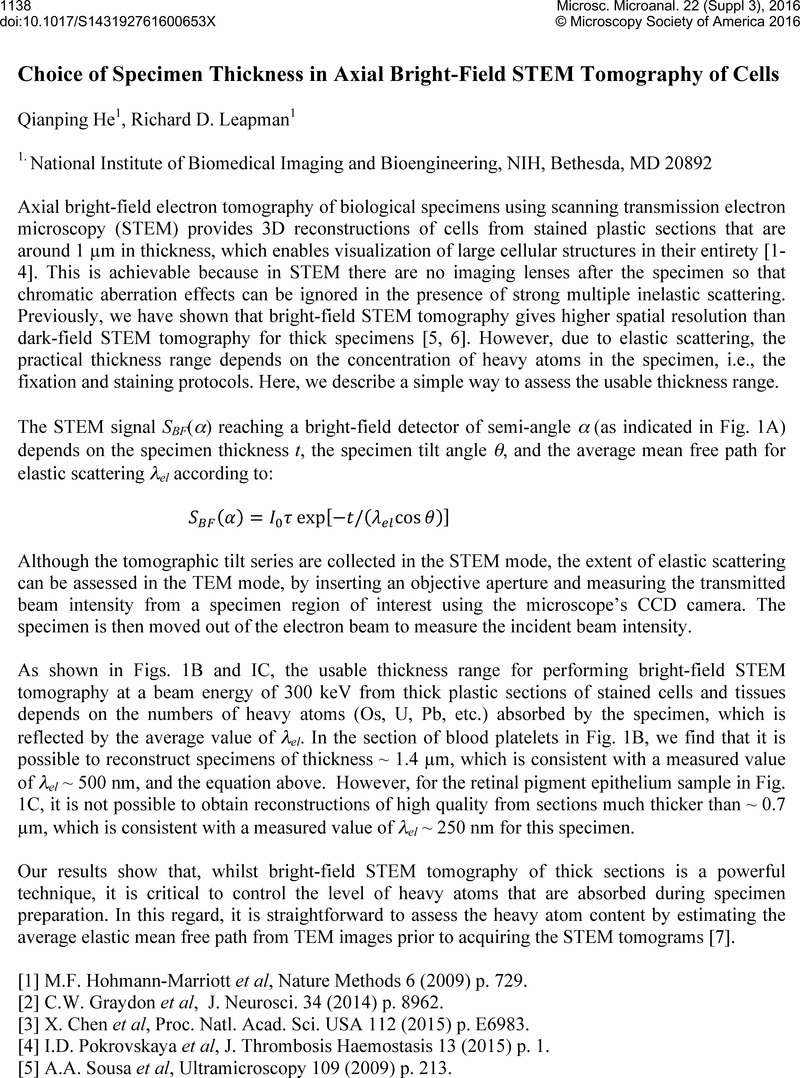
Image of the first page of this content. For PDF version, please use the ‘Save PDF’ preceeding this image.'
Type
Abstract
Copyright
© Microscopy Society of America 2016 

References

[1] Hohmann-Marriott, M.F., et al, Nature Methods 6 (2009). p. 729.Google Scholar
[2] Graydon, C.W., et al, J. Neurosci 34 (2014). p. 8962.Google Scholar
[3] Chen, X., et al, Proc. Natl. Acad. Sci. USA 112 (2015). p. E6983.Google Scholar
[4] Pokrovskaya, I.D., et al, J. Thrombosis Haemostasis 13 (2015). p. 1.Google Scholar
[5] Sousa, A.A., et al, Ultramicroscopy 109 (2009). p. 213.Google Scholar
[6] Sousa, A.A., et al, J. Struct. Biol 174 (2011). p. 107.Google Scholar
[7] The authors thank Drs. B. Storrie and G. Zhang for providing the blood platelet specimen, and Drs. Q. Lu and C.J. Westlake for the providing the retinal pigment epithelium specimen. This research was supported by the intramural program of the National Institute of Biomedical Imaging and Bioengineering, NIH.Google Scholar