Hostname: page-component-586b7cd67f-dlnhk Total loading time: 0 Render date: 2024-11-23T14:47:31.443Z Has data issue: false hasContentIssue false

Analysis of Electron Channeling Contrast of Stacking Faults in fcc Materials

Published online by Cambridge University Press:  22 January 2021
Ivan Gutierrez-Urrutia*
Affiliation:
Research Center for Strategic Materials, National Institute for Materials Science, Tsukuba, Ibaraki305-0047, Japan
*
Author for correspondence: Ivan Gutierrez-Urrutia, E-mail: [email protected]
Get access

Abstract

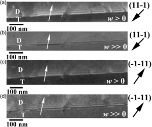
The characteristics of electron channeling contrast (ECC) of stacking faults in a [101] single-crystal 316L fcc (face-centered cubic) stainless steel have been evaluated. Channeling contrast was analyzed from a series of ECC images taken as a function of sample tilt at ~0.1° increments across the (111) Kikuchi band. The most relevant imaging parameters of the fault contrast, namely the number of fringes, fringe intensity, and fringe spacing, were analyzed under different channeling conditions. The present work shows that the channeling contrast of stacking faults exhibits strong dependence on the sign and magnitude of the deviation parameter, w (w = s ξg, where s is the excitation vector and ξg is the extinction distance). This effect has strong influence on channeling conditions for fault imaging and g.R analysis for determining the nature of a stacking fault. g.R analysis was evaluated by the method developed by Gevers et al. (1963) on g-reversion experiments of ECC images of a stacking fault configuration. The interplay between the stacking fault nature and ε-martensite is analyzed.

Type
Software and Instrumentation
Copyright
Copyright © The Author(s), 2021. Published by Cambridge University Press on behalf of the Microscopy Society of America

Access options

Get access to the full version of this content by using one of the access options below. (Log in options will check for institutional or personal access. Content may require purchase if you do not have access.)

References

Anderson, PM, Hirth, JP & Lothe, J (2017). Theory of Dislocations. Cambridge: Cambridge University Press.Google Scholar
Art, E, Gevers, R & Amelinckx, S (1963). The determination of the type of stacking faults in face centered cubic alloys by means of contrast effects in the electron microscope. Phys Status Solidi 3, 697711.CrossRefGoogle Scholar
Bithell, EG, Donovan, PE & Stobbs, WM (1989). The relative effects of specimen thickness and convergence on the weakbeam contrast of stacking faults. Philos Mag A 59, 6385.CrossRefGoogle Scholar
Booker, GR & Hazzledine, PM (1967). Electron microscope image profiles of planar defects in crystals. Philos Mag 15, 523527.CrossRefGoogle Scholar
Brooks, JW, Loretto, MH & Smallman, RE (1979 a). Direct observations of martensite nuclei in stainless steel. Acta Metall 27, 18391847.CrossRefGoogle Scholar
Brooks, JW, Loretto, MH & Smallman, RE (1979 b). In situ observations of the formation of martensite in stainless steel. Acta Metall 27, 18291838.CrossRefGoogle Scholar
Crimp, MA, Simkin, BA & Ng, BC (2001). Demonstration of the g⋅ b x u=0 edge dislocation invisibility criterion for electron channeling contrast imaging. Philos Mag Lett 81, 833837.CrossRefGoogle Scholar
Cullis, AG & Booker, GR (1972). Proc. Fifth European Conference on Electron Microscopy, Manchester. London. The Institute of Physics, 532–533.Google Scholar
Curtze, S, Kuokkala, V-T, Oikari, A, Talonen, J & Hänninen, H (2011). Thermodynamic modeling of the stacking fault energy of austenitic steels. Acta Mater 59, 10681076.CrossRefGoogle Scholar
Dash, J & Otte, HM (1963). The martensite transformation in stainless steel. Acta Metall 11, 11691178.CrossRefGoogle Scholar
Dunlap, BE, Ruggles, TJ, Fullwood, DT, Jackson, B & Crimp, MA (2018). Comparison of dislocation characterization by electron channeling contrast imaging and cross-correlation electron backscattered diffraction. Ultramicroscopy 184, 125133.CrossRefGoogle ScholarPubMed
Feifel, M, Ohlmann, J, France, RM, Lackner, D & Dimroth, F (2020). Electron channeling contrast imaging investigation of stacking fault pyramids in GaP on Si nucleation layers. J Crystal Growth 532, 125422.CrossRefGoogle Scholar
Föll, H, Carter, CB & Wilkens, M (1980). Weak-beam contrast of stacking faults in transmission electron microscopy. Phys Status Solidi A 58, 393407.CrossRefGoogle Scholar
Frank, FC (1951). Crystal dislocations—Elementary concepts and definitions. Philos Mag 42, 809819.CrossRefGoogle Scholar
Fujita, H & Ueda, S (1972). Stacking faults and fcc (γ) → hcp (ϵ) transformation in 18/8-type stainless steel. Acta Metall 20, 759767.CrossRefGoogle Scholar
Fultz, B & Howe, JM (2013). Transmission Electron Microscopy and Diffractometry of Materials. Berlin: Springer-Verlag.CrossRefGoogle Scholar
Gevers, R, Art, A & Amelinckx, S (1963). Electron microscopic images of single and intersecting stacking faults in thick foils. Part I: Single faults. Phys Status Solidi 3, 15631593.CrossRefGoogle Scholar
Gutierrez-Urrutia, I (2017). Analysis of FIB induced damage by electron channeling contrast imaging in the SEM. J Microsc 265, 5159.CrossRefGoogle Scholar
Gutierrez-Urrutia, I (2019). Quantitative analysis of electron channeling contrast of dislocations. Ultramicroscopy 206, 112826.CrossRefGoogle ScholarPubMed
Gutierrez-Urrutia, I, Zaefferer, S & Raabe, D (2013). Coupling of electron channeling with EBSD: Toward the quantitative characterization of deformation structures in the SEM. JOM 65, 12291236.CrossRefGoogle Scholar
Hashimoto, H (1964). Energy dependence of extinction distance and transmissive power for electron waves in crystals. J Appl Phys 35, 277290.CrossRefGoogle Scholar
Hashimoto, H, Howie, A & Whelan, MJ (1962). Anomalous electron absorption effects in metal foils: Theory and comparison with experiment. Proc Phys Soc 269, 80103.Google Scholar
Hirsch, P, Howie, A, Nicholson, RB, Pashley, DW & Whelan, MJ (1965). Electron Microscopy of Thin Crystals, 1st ed. London: Butterworths.Google Scholar
Howie, A & Valdre, U (1963). The nature of deformation stacking faults in F.C.C. alloys. Philos Mag 8, 19811984.CrossRefGoogle Scholar
Howie, A & Whelan, MJ (1962). Diffraction contrast of electron microscope images of crystal lattice defects. III. Results and experimental confirmation of the dynamical theory of dislocation image contrast. Proc R Soc A 267, 206230.Google Scholar
Humphreys, CJ, Howie, A & Booker, GR (1967). Some electron diffraction contrast effects at planar defects in crystals. Philos Mag 15, 507522.CrossRefGoogle Scholar
Kelly, PM (1965). The martensite transformation in steels with low stacking fault energy. Acta Metall 13, 635646.CrossRefGoogle Scholar
Kriaa, H, Guitton, A & Maloufi, N (2017). Fundamental and experimental aspects of diffraction for characterizing dislocations by electron channeling contrast imaging in scanning electron microscope. Sci Rep 7, 9742.CrossRefGoogle ScholarPubMed
Kriaa, H, Guitton, A & Maloufi, N (2019). Modeling dislocation contrasts obtained by accurate-electron channeling contrast imaging for characterizing deformation mechanisms in bulk materials. Materials 12, 1587.CrossRefGoogle ScholarPubMed
Mahajan, S, Green, ML & Brasen, D (1977). A model for the FCC→HCP transformation, its applications, and experimental evidence. Metall Trans A 8A, 283293.CrossRefGoogle Scholar
Makineni, SK, Kumar, A, Lenz, M, Kontis, P, Meiners, T, Zenk, C & Gault, B (2018). On the diffusive phase transformation mechanism assisted by extended dislocations during creep of a single crystal CoNi-based superalloy. Acta Mater 155, 362371.CrossRefGoogle Scholar
Morin, P, Pitaval, M, Besnard, D & Fontaine, G (1979). Electron-channelling imaging in scanning electron microscopy. Philos Mag A 40, 511524.CrossRefGoogle Scholar
Morniroli, JP (2006). Electron Diffraction Version 7.2, Dedicated Software to Interpret LACBED Patterns. Lille: USTL.Google Scholar
Naresh-Kumar, G, Thomson, D, Zhang, Y, Bai, J, Jiu, L, Yu, X & Trager-Cowan, C (2018). Imaging basal plane stacking faults and dislocations in (11-22) GaN using electron channeling contrast imaging. J Appl Phys 124, 065301.CrossRefGoogle Scholar
Olson, GB & Cohen, M (1975). Kinetics of nucleation strain-induced martensitic. Metall Trans A 6A, 791795.CrossRefGoogle Scholar
Olson, GB & Cohen, M (1976). A general mechanism of martensitic nucleation: Part I. General concepts and the FCC→HCP transformation. Metall Trans A 7A, 18971904.Google Scholar
Olson, GB & Cohen, M (1981). A perspective on martensitic nucleation. Ann Rev Mater Sci 11, 130.CrossRefGoogle Scholar
Pang, B, Jones, IP, Chiu, Y-L, Millett, JCF & Whiteman, G (2017). Electron channeling contrast imaging of dislocations in a conventional SEM. Philos Mag 97, 346359.CrossRefGoogle Scholar
Picard, YN, Liu, M, Lammatao, J, Kamaladasa, R & Graef, MD (2014). Theory of dynamical electron channeling contrast images of near-surface crystal defects. Ultramicroscopy 146, 7178.CrossRefGoogle ScholarPubMed
Reimer, L & Kohl, H (2008). Transmission Electron Microscopy. Physics of Image Formation, 5th ed. Berlin: Springer.Google Scholar
Simkin, BA & Crimp, MA (1999). An experimentally convenient configuration for electron channeling contrast imaging. Ultramicroscopy 77, 6575.CrossRefGoogle Scholar
Spencer, JP, Humphreys, CJ & Hirsch, PB (1972). A dynamical theory for the contrast of perfect and imperfect crystals in the scanning electron microscope using backscattered electrons. Philos Mag 26, 193213.CrossRefGoogle Scholar
Talonen, J & Hänninen, H (2007). Formation of shear bands and strain-induced martensite during plastic deformation of metastable austenitic stainless steels. Acta Mater 55, 61086118.CrossRefGoogle Scholar
Trager-Cowan, C, Sweeney, F, Trimby, PW, Day, AP, Gholinia, A, Schmidt, NH & Watson, IM (2007). Electron backscatter diffraction and electron channeling contrast imaging of tilt and dislocations in nitride thin films. Phys Rev B 75, 085301.CrossRefGoogle Scholar
Twigg, ME & Picard, YN (2009). Simulation and analysis of electron channeling contrast images of threading screw dislocations in 4H-SiC. J Appl Phys 105, 093520.CrossRefGoogle Scholar
Venables, JA (1962). The martensite transformation in stainless steel. Philos Mag 7(73), 3544.CrossRefGoogle Scholar
Vitos, L, Nilsson, J-O & Johansson, B (2006). Alloying effects on the stacking fault energy in austenitic stainless steels from first-principles theory. Acta Mater 54, 38213826.CrossRefGoogle Scholar
Wang, M, Li, Z & Raabe, D (2018). In-situ SEM observation of phase transformation and twinning mechanisms in an interstitial high-entropy alloy. Acta Mater 147, 236246.CrossRefGoogle Scholar
Weidner, A & Biermann, H (2017). Case studies on the application of high-resolution electron channeling contrast imaging—Investigation of defects and defect arrangements in metallic materials. Philos Mag 95, 759793.CrossRefGoogle Scholar
Weidner, A, Martin, S, Klemm, V, Martin, U & Biermann, H (2011). Stacking faults in high-alloyed metastable austenitic cast steel observed by electron channeling contrast imaging. Scr Mater 64, 513516.CrossRefGoogle Scholar
Whelan, MJ & Hirsch, PB (1957a). Electron diffraction from crystals containing stacking faults: I. Philos Mag 2(21), 11211142.CrossRefGoogle Scholar
Whelan, MJ & Hirsch, PB (1957b). Electron diffraction from crystals containing stacking faults: II. Philos Mag 2(23), 13031324.CrossRefGoogle Scholar
Wilkinson, AJ, Anstis, GR, Czernuszka, JT, Long, NJ & Hirsch, PB (1993). Electron channeling contrast imaging of interfacial defects in strained silicon-germanium layers on silicon. Philos Mag A 68, 5980.CrossRefGoogle Scholar
Wilkinson, AJ & Hirsch, PB (1997). Electron diffraction based techniques in scanning electron microscopy of bulk materials. Micron 28(4), 279308.CrossRefGoogle Scholar
Williams, DB & Carter, CB (2009). Transmission Electron Microscopy. A Textbook for Materials Science. New York: Springer.CrossRefGoogle Scholar
Yang, X-S, Sun, S & Zhang, T-Y (2015). The mechanism of bcc α′ nucleation in single hcp ε laths in the fcc γ → hcp ε → bcc α′ martensitic phase transformation. Acta Mater 95, 264273.CrossRefGoogle Scholar
Zaefferer, S & Elhami, N-N (2014). Overview No. 154. Theory and application of electron channeling contrast imaging under controlled diffraction conditions. Acta Mater 75, 2050.CrossRefGoogle Scholar
Zauter, R, Petry, F, Bayerlein, M, Sommer, C, Christ, HJ & Mughrabi, H (1992). Electron channeling contrast as a supplementary method for microstructural investigations in deformed metals. Philos Mag A 66, 425436.CrossRefGoogle Scholar