Hostname: page-component-586b7cd67f-vdxz6 Total loading time: 0 Render date: 2024-11-22T11:22:48.109Z Has data issue: false hasContentIssue false

Accurate and Robust Calibration of the Uniform Affine Transformation Between Scan-Camera Coordinates for Atom-Resolved In-Focus 4D-STEM Datasets

Published online by Cambridge University Press:  09 March 2022
Shoucong Ning
Affiliation:
Department of Materials Science and Engineering, National University of Singapore, Singapore 117575, Singapore Center for Bio-Imaging Sciences, National University of Singapore, Singapore 117557, Singapore
Wenhui Xu
Affiliation:
Department of Electrical and Electronic Engineering, Southern University of Science and Technology, Shenzhen 518055, China Harbin Institute of Technology, Harbin 150001, China
Yinhang Ma
Affiliation:
School of Physical Sciences and CAS Key Laboratory of Vacuum Physics, University of Chinese Academy of Sciences, Beijing 100049, China
Leyi Loh
Affiliation:
Department of Materials Science and Engineering, National University of Singapore, Singapore 117575, Singapore
Timothy J. Pennycook
Affiliation:
EMAT, University of Antwerp, Campus Groenenborger, 2020 Antwerp, Belgium
Wu Zhou
Affiliation:
School of Physical Sciences and CAS Key Laboratory of Vacuum Physics, University of Chinese Academy of Sciences, Beijing 100049, China
Fucai Zhang
Affiliation:
Department of Electrical and Electronic Engineering, Southern University of Science and Technology, Shenzhen 518055, China
Michel Bosman
Affiliation:
Department of Materials Science and Engineering, National University of Singapore, Singapore 117575, Singapore
Stephen J. Pennycook
Affiliation:
School of Physical Sciences and CAS Key Laboratory of Vacuum Physics, University of Chinese Academy of Sciences, Beijing 100049, China
Qian He*
Affiliation:
Department of Materials Science and Engineering, National University of Singapore, Singapore 117575, Singapore
N. Duane Loh*
Affiliation:
Center for Bio-Imaging Sciences, National University of Singapore, Singapore 117557, Singapore Department of Physics, National University of Singapore, Singapore 117551, Singapore Department of Biological Sciences, National University of Singapore, Singapore 117557, Singapore
*
*Corresponding authors: Qian He, E-mail: [email protected]; N. Duane Loh, E-mail: [email protected]
Get access

Abstract

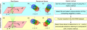
Accurate geometrical calibration between the scan coordinates and the camera coordinates is critical in four-dimensional scanning transmission electron microscopy (4D-STEM) for both quantitative imaging and ptychographic reconstructions. For atomic-resolved, in-focus 4D-STEM datasets, we propose a hybrid method incorporating two sub-routines, namely a J-matrix method and a Fourier method, which can calibrate the uniform affine transformation between the scan-camera coordinates using raw data, without a priori knowledge of the crystal structure of the specimen. The hybrid method is found robust against scan distortions and residual probe aberrations. It is also effective even when defects are present in the specimen, or the specimen becomes relatively thick. We will demonstrate that a successful geometrical calibration with the hybrid method will lead to a more reliable recovery of both the specimen and the electron probe in a ptychographic reconstruction. We will also show that, although the elimination of local scan position errors still requires an iterative approach, the rate of convergence can be improved, and the residual errors can be further reduced if the hybrid method can be firstly applied for initial calibration. The code is made available as a simple-to-use tool to correct affine transformations of the scan-camera coordinates in 4D-STEM experiments.

Type
Software and Instrumentation
Copyright
Copyright © The Author(s), 2022. Published by Cambridge University Press on behalf of the Microscopy Society of America

Access options

Get access to the full version of this content by using one of the access options below. (Log in options will check for institutional or personal access. Content may require purchase if you do not have access.)

References

Addiego, C, Gao, W & Pan, X (2020). Thickness and defocus dependence of inter-atomic electric fields measured by scanning diffraction. Ultramicroscopy 208, 112850.CrossRefGoogle ScholarPubMed
Ballabriga, R, Campbell, M, Heijne, E, Llopart, X, Tlustos, L & Wong, W (2011). Medipix3: A 64k pixel detector readout chip working in single photon counting mode with improved spectrometric performance. Nucl Instrum Methods Phys Res A 633, S15S18.CrossRefGoogle Scholar
Berkels, B, Binev, P, Blom, DA, Dahmen, W, Sharpley, RC & Vogt, T (2014). Optimized imaging using non-rigid registration. Ultramicroscopy 138, 4656.CrossRefGoogle ScholarPubMed
Berkels, B & Liebscher, CH (2019). Joint non-rigid image registration and reconstruction for quantitative atomic resolution scanning transmission electron microscopy. Ultramicroscopy 198, 4957.CrossRefGoogle ScholarPubMed
Chen, Z, Odstrcil, M, Jiang, Y, Han, Y, Chiu, M-H, Li, L-J & Muller, DA (2020). Mixed-state electron ptychography enables sub-angstrom resolution imaging with picometer precision at low dose. Nat Commun 11, 2994.CrossRefGoogle ScholarPubMed
Ciston, J, Johnson, IJ, Draney, BR, Ercius, P, Fong, E, Goldschmidt, A, Joseph, JM, Lee, JR, Mueller, A, Ophus, C, Selvarajan, A, Skinner, DE, Stezelberger, T, Tindall, CS, Minor, AM & Denes, P (2019). The 4D camera: Very high speed electron counting for 4D-STEM. Microsc Microanal 25, 19301931.CrossRefGoogle Scholar
Dwivedi, P, Konijnenberg, AP, Pereira, SF & Urbach, HP (2018). Lateral position correction in ptychography using the gradient of intensity patterns. Ultramicroscopy 192, 2936.CrossRefGoogle ScholarPubMed
Ercius, P, Johnson, I, Brown, H, Pelz, P, Hsu, S-L, Draney, B, Fong, E, Goldschmidt, A, Joseph, J, Lee, J, Ciston, J, Ophus, C, Scott, M, Selvarajan, A, Paul, D, Skinner, D, Hanwell, M, Harris, C, Avery, P, Stezelberger, T, Tindall, C, Ramesh, R, Minor, A & Denes, P (2020). The 4D camera – An 87 kHz frame-rate detector for counted 4D-STEM experiments. Microsc Microanal 26, 18961897.CrossRefGoogle Scholar
Gao, W, Addiego, C, Wang, H, Yan, X, Hou, Y, Ji, D, Heikes, C, Zhang, Y, Li, L, Huyan, H, Blum, T, Aoki, T, Nie, Y, Schlom, DG, Wu, R & Pan, X (2019). Real-space charge-density imaging with sub-ångström resolution by four-dimensional electron microscopy. Nature 575, 480484.CrossRefGoogle ScholarPubMed
Guizar-Sicairos, M & Fienup, JR (2008). Phase retrieval with transverse translation diversity: A nonlinear optimization approach. Optics Express 16, 72647278.CrossRefGoogle ScholarPubMed
Haas, B, Schloz, M, Mittelberger, A, Lovejoy, T, Müller, J, Krivanek, O, Jones, L, Van den Broek, W & Koch, C (2020). Comparison of ptychography vs. center-of-mass analysis of registered 4D-STEM series. Microsc Microanal 26, 18981900.CrossRefGoogle Scholar
Hachtel, JA, Idrobo, JC & Chi, M (2018). Sub-Ångstrom electric field measurements on a universal detector in a scanning transmission electron microscope. Adv Struct Chem Imaging 4, 10.CrossRefGoogle Scholar
Heimes, D, Belz, J, Beyer, A & Volz, K (2020). Measuring interatomic bonding and charge redistributions in defects by combining 4D-STEM and STEM multislice simulations. Microsc Microanal 26, 452454.CrossRefGoogle Scholar
Jannis, D, Hofer, C, Gao, C, Gao, X, Béché, A, Pennycook, TJ & Verbeeck, J (2021). Event driven 4D STEM acquisition with a Timepix3 detector: Microsecond dwell time and faster scans for high precision and low dose applications. Ultramicroscopy 233, 113423.CrossRefGoogle ScholarPubMed
Jiang, Y, Chen, Z, Han, Y, Deb, P, Gao, H, Xie, S, Purohit, P, Tate, MW, Park, J, Gruner, SM, Elser, V & Muller, DA (2018). Electron ptychography of 2D materials to deep sub-ångström resolution. Nature 559, 343349.CrossRefGoogle Scholar
Jones, L (2014). Scan-noise and drift correction in the STEM. Microsc Today 22, 4041.CrossRefGoogle Scholar
Jones, L & Nellist, PD (2013). Identifying and correcting scan noise and drift in the scanning transmission electron microscope. Microsc Microanal 19, 10501060.CrossRefGoogle ScholarPubMed
Jones, L, Wenner, S, Nord, M, Ninive, PH, Løvvik, OM, Holmestad, R & Nellist, PD (2017). Optimising multi-frame ADF-STEM for high-precision atomic-resolution strain mapping. Ultramicroscopy 179, 5762.CrossRefGoogle ScholarPubMed
Lazić, I, Bosch, EGT & Lazar, S (2016). Phase contrast STEM for thin samples: Integrated differential phase contrast. Ultramicroscopy 160, 265280.CrossRefGoogle ScholarPubMed
Maiden, AM, Humphry, MJ, Sarahan, MC, Kraus, B & Rodenburg, JM (2012). An annealing algorithm to correct positioning errors in ptychography. Ultramicroscopy 120, 6472.CrossRefGoogle ScholarPubMed
Maiden, AM & Rodenburg, JM (2009). An improved ptychographical phase retrieval algorithm for diffractive imaging. Ultramicroscopy 109, 12561262.CrossRefGoogle ScholarPubMed
Müller, K, Krause, FF, Béché, A, Schowalter, M, Galioit, V, Löffler, S, Verbeeck, J, Zweck, J, Schattschneider, P & Rosenauer, A (2014). Atomic electric fields revealed by a quantum mechanical approach to electron picodiffraction. Nat Commun 5, 5653.CrossRefGoogle ScholarPubMed
Müller-Caspary, K, Grieb, T, Müßener, J, Gauquelin, N, Hille, P, Schörmann, J, Verbeeck, J, Van Aert, S, Eickhoff, M & Rosenauer, A (2019). Electrical polarization in AlN/GaN nanodisks measured by momentum-resolved 4D scanning transmission electron microscopy. Phys Rev Lett 122, 106102.CrossRefGoogle ScholarPubMed
Nguyen, KX, Purohit, P, Hovden, R, Turgut, E, Tate, MW, Kourkoutis, LF, Fuchs, GD, Gruner, SM & Muller, DA (2016). 4D-STEM for quantitative imaging of magnetic materials with enhanced contrast and resolution. Microsc Microanal 22, 17181719.CrossRefGoogle Scholar
Ning, S, Fujita, T, Nie, A, Wang, Z, Xu, X, Chen, J, Chen, M, Yao, S & Zhang, T-Y (2018). Scanning distortion correction in STEM images. Ultramicroscopy 184, 274283.CrossRefGoogle ScholarPubMed
Nord, M, Webster, RWH, Paton, KA, McVitie, S, McGrouther, D, MacLaren, I & Paterson, GW (2020). Fast pixelated detectors in scanning transmission electron microscopy. Part I: Data acquisition, live processing, and storage. Microsc Microanal 26, 653666.CrossRefGoogle ScholarPubMed
O'Leary, C, Haas, B, Koch, C, Nellist, P & Jones, L (2021). Increasing spatial fidelity and SNR of 4D-STEM using multi-frame data fusion. Microsc Microanal, 111. https://doi.org/10.1017/S1431927621012587.Google Scholar
O'Leary, CM, Allen, CS, Huang, C, Kim, JS, Liberti, E, Nellist, PD & Kirkland, AI (2020). Phase reconstruction using fast binary 4D STEM data. Appl Phys Lett 116, 124101.CrossRefGoogle Scholar
Ophus, C (2019). Four-Dimensional scanning transmission electron microscopy (4D-STEM): From scanning nanodiffraction to ptychography and beyond. Microsc Microanal 25, 563582.CrossRefGoogle ScholarPubMed
Ophus, C, Ercius, P, Sarahan, M, Czarnik, C & Ciston, J (2014). Recording and using 4D-STEM datasets in materials science. Microsc Microanal 20, 6263.CrossRefGoogle Scholar
Pelz, PM, Qiu, WX, Bücker, R, Kassier, G & Miller, RJD (2017). Low-dose cryo electron ptychography via non-convex Bayesian optimization. Sci Rep 7, 9883.CrossRefGoogle ScholarPubMed
Pennycook, SJ (2017). The impact of STEM aberration correction on materials science. Ultramicroscopy 180, 2233.CrossRefGoogle ScholarPubMed
Pennycook, TJ, Lupini, AR, Yang, H, Murfitt, MF, Jones, L & Nellist, PD (2015). Efficient phase contrast imaging in STEM using a pixelated detector. Part 1: Experimental demonstration at atomic resolution. Ultramicroscopy 151, 160167.CrossRefGoogle ScholarPubMed
Pennycook, TJ, Martinez, GT, Nellist, PD & Meyer, JC (2019). High dose efficiency atomic resolution imaging via electron ptychography. Ultramicroscopy 196, 131135.CrossRefGoogle ScholarPubMed
Plackett, R, Horswell, I, Gimenez, EN, Marchal, J, Omar, D & Tartoni, N (2013). Merlin: A fast versatile readout system for Medipix3. J Instrum 8, C01038.CrossRefGoogle Scholar
Rodenburg, J & Maiden, A (2019). Ptychography. Springer Handbook of Microscopy.CrossRefGoogle Scholar
Savitzky, BH, Zeltmann, SE, Hughes, LA, Brown, HG, Zhao, S, Pelz, PM, Pekin, TC, Barnard, ES, Donohue, J, Rangel DaCosta, L, Kennedy, E, Xie, Y, Janish, MT, Schneider, MM, Herring, P, Gopal, C, Anapolsky, A, Dhall, R, Bustillo, KC, Ercius, P, Scott, MC, Ciston, J, Minor, AM & Ophus, C (2021). py4DSTEM: A software package for four-dimensional scanning transmission electron microscopy data analysis. Microsc Microanal 27, 712743.CrossRefGoogle ScholarPubMed
Shenfield, A & Rodenburg, JM (2011). Evolutionary determination of experimental parameters for ptychographical imaging. J Appl Phys 109, 124510.CrossRefGoogle Scholar
Tinti, G, Fröjdh, E, van Genderen, E, Gruene, T, Schmitt, B, de Winter, DAM, Weckhuysen, BM & Abrahams, JP (2018). Electron crystallography with the EIGER detector. IUCrJ 5, 190199.CrossRefGoogle ScholarPubMed
Wen, Y, Ophus, C, Allen, CS, Fang, S, Chen, J, Kaxiras, E, Kirkland, AI & Warner, JH (2019). Simultaneous identification of low and high atomic number atoms in monolayer 2D materials using 4D scanning transmission electron microscopy. Nano Lett 19, 64826491.CrossRefGoogle Scholar
Xu, W, Lin, H, Wang, H & Zhang, F (2020). Reconstruction method of a ptychographic dataset with unknown positions. Opt Lett 45, 46344637.CrossRefGoogle ScholarPubMed
Yang, H, Jones, L, Ryll, H, Simson, M, Soltau, H, Kondo, Y, Sagawa, R, Banba, H, MacLaren, I & Nellist, PD (2015 a). 4D STEM: High efficiency phase contrast imaging using a fast pixelated detector. J Phys Conf Series 644, 012032.CrossRefGoogle Scholar
Yang, H, Pennycook, TJ & Nellist, PD (2015 b). Efficient phase contrast imaging in STEM using a pixelated detector. Part II: Optimisation of imaging conditions. Ultramicroscopy 151, 232239.CrossRefGoogle ScholarPubMed
Yang, H, Rutte, RN, Jones, L, Simson, M, Sagawa, R, Ryll, H, Huth, M, Pennycook, TJ, Green, MLH, Soltau, H, Kondo, Y, Davis, BG & Nellist, PD (2016). Simultaneous atomic-resolution electron ptychography and Z-contrast imaging of light and heavy elements in complex nanostructures. Nat Commun 7, 12532.CrossRefGoogle ScholarPubMed
Zhang, F, Peterson, I, Vila-Comamala, J, Diaz, A, Berenguer, F, Bean, R, Chen, B, Menzel, A, Robinson, IK & Rodenburg, JM (2013). Translation position determination in ptychographic coherent diffraction imaging. Opt Express 21, 1359213606.CrossRefGoogle ScholarPubMed
Zhou, L, Song, J, Kim, JS, Pei, X, Huang, C, Boyce, M, Mendonça, L, Clare, D, Siebert, A, Allen, CS, Liberti, E, Stuart, D, Pan, X, Nellist, PD, Zhang, P, Kirkland, AI & Wang, P (2020). Low-dose phase retrieval of biological specimens using cryo-electron ptychography. Nat Commun 11, 2773.CrossRefGoogle ScholarPubMed
Supplementary material: File

Ning et al. supplementary material

Ning et al. supplementary material

Download Ning et al. supplementary material(File)
File 10.2 MB