Hostname: page-component-586b7cd67f-g8jcs Total loading time: 0 Render date: 2024-11-26T06:14:55.305Z Has data issue: false hasContentIssue false

Three-Dimensional Analysis of Interstitial Cells in the Smooth Muscle Layer of Murine Vas Deferens Using Confocal Laser Scanning Microscopy and FIB/SEM

Published online by Cambridge University Press:  26 January 2022

Tasuku Hiroshige*
Affiliation:
Department of Urology, Kurume University School of Medicine, Kurume 830-0011, Japan Division of Microscopic and Development Anatomy, Department of Anatomy, Kurume University School of Medicine, Kurume 830-0011, Japan
Kei-Ichiro Uemura
Affiliation:
Department of Urology, Kurume University School of Medicine, Kurume 830-0011, Japan
Shingo Hirashima
Affiliation:
Division of Microscopic and Development Anatomy, Department of Anatomy, Kurume University School of Medicine, Kurume 830-0011, Japan
Kiyosato Hino
Affiliation:
Division of Microscopic and Development Anatomy, Department of Anatomy, Kurume University School of Medicine, Kurume 830-0011, Japan
Akinobu Togo
Affiliation:
Advanced Imaging Research Center, Kurume University School of Medicine, Kurume 830-0011, Japan
Keisuke Ohta
Affiliation:
Division of Microscopic and Development Anatomy, Department of Anatomy, Kurume University School of Medicine, Kurume 830-0011, Japan Advanced Imaging Research Center, Kurume University School of Medicine, Kurume 830-0011, Japan
Tsukasa Igawa
Affiliation:
Department of Urology, Kurume University School of Medicine, Kurume 830-0011, Japan
Kei-Ichiro Nakamura
Affiliation:
Division of Microscopic and Development Anatomy, Department of Anatomy, Kurume University School of Medicine, Kurume 830-0011, Japan Cognitive and Molecular Research Institute of Brain Diseases, Kurume University School of Medicine, Kurume 830-0011, Japan
*
*Corresponding author: Tasuku Hiroshige, E-mail: [email protected]
Get access

Abstract

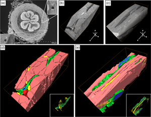
The smooth muscle contraction of the vas deferens has the important function of transporting sperm. Interstitial cells (ICs) play a critical role in the pacing and modulation of various smooth muscle organs by interactions with nerves and smooth muscle. Elucidating the three-dimensional (3D) architecture of ICs is important for understanding their spatial relationship on the mesoscale between ICs, smooth muscle cells (SMCs), and nerves. In this study, the 3D ultrastructure of ICs in the smooth muscle layer of murine vas deferens and the spatial relationships between ICs, nerves, and smooth muscles were observed using confocal laser scanning microscopy and focused ion beam/scanning electron microscopy. ICs have sheet-like structures as demonstrated by 3D observation using modern analytical techniques. Sheet-like ICs have two types of 3D structures, one flattened and the other curled. Multiple extracellular vesicle (EV)-like structures were frequently observed in ICs. Various spatial relations were observed in areas between ICs, nerves, and SMCs, which formed a complex 3D network with each other. These results suggest that ICs in the smooth muscle layer of murine vas deferens may have two subtypes with different sheet-like structures and may be involved in neuromuscular signal transmission via physical interaction and EVs.

Type
Micrographia
Copyright
Copyright © The Author(s), 2022. Published by Cambridge University Press on behalf of the Microscopy Society of America

Access options

Get access to the full version of this content by using one of the access options below. (Log in options will check for institutional or personal access. Content may require purchase if you do not have access.)

References

Amelar, RD, Dubin, L & Schoenfeld, C (1980). Sperm motility. Fertil Steril 34, 197215. doi:10.1016/s0015-0282(16)44949-6Google ScholarPubMed
Andrae, J, Gallini, R & Betsholtz, C (2008). Role of platelet-derived growth factors in physiology and medicine. Genes Dev 22, 12761312.CrossRefGoogle ScholarPubMed
Cretoiu, D (2016). The third dimension of telocytes revealed by FIB-SEM tomography. Adv Exp Med Biol 913, 325334. doi:10.1007/978-981-10-1061-3_21CrossRefGoogle ScholarPubMed
Cretoiu, D, Gherghiceanu, M, Hummel, E, Zimmermann, H, Simionescu, O & Popescu, LM (2015). FIB-SEM tomography of human skin telocytes and their extracellular vesicles. J Cell Mol Med 19, 714722. doi:10.1111/jcmm.12578CrossRefGoogle ScholarPubMed
Cretoiu, D, Hummel, E, Zimmermann, H, Gherghiceanu, M & Popescu, LM (2014). Human cardiac telocytes: 3D imaging by FIB-SEM tomography. J Cell Mol Med 18, 21572164. doi:10.1111/jcmm.12468CrossRefGoogle ScholarPubMed
Cretoiu, SM & Popescu, LM (2014). Telocytes revisited. Biomol Concepts 5, 353369. doi:10.1515/bmc-2014-0029CrossRefGoogle ScholarPubMed
Edelstein, L & Smythies, J (2014). The role of telocytes in morphogenetic bioelectrical signaling: Once more unto the breach. Front Mol Neurosci 7, 41. doi:10.3389/fnmol.2014.00041.CrossRefGoogle ScholarPubMed
Gherghiceanu, M & Popescu, LM (2012). Cardiac telocytes – their junctions and functional implications. Cell Tissue Res 348, 265279. doi:10.1007/s00441-012-1333-8CrossRefGoogle ScholarPubMed
Hashitani, H, Nguyen, MJ, Noda, H, Mitsui, R, Higashi, R, Ohta, K, Nakamura, K-I & Lang, RJ (2017). Interstitial cell modulation of pyeloureteric peristalsis in the mouse renal pelvis examined using FIBSEM tomography and calcium indicators. Pflugers Arch 469, 797813. doi:10.1007/s00424-016-1930-6CrossRefGoogle ScholarPubMed
Hiroshige, T, Uemura, K-I, Hirashima, S, Hino, K, Togo, A, Ohta, K, Igawa, T & Nakamura, K-I (2021). Identification of PDGFRα-positive interstitial cells in the distal segment of the murine vas deferens. Sci Rep 11, 7553. doi:10.1038/s41598-021-87049-6.CrossRefGoogle ScholarPubMed
Kizilyaprak, C, Stierhof, YD & Humbel, BM (2019). Volume microscopy in biology: FIB-SEM tomography. Tissue Cell 57, 123128. doi:10.1016/j.tice.2018.09.006CrossRefGoogle ScholarPubMed
Koh, SD, Lee, H, Ward, SM & Sanders, KM (2018). The mystery of the interstitial cells in the urinary bladder. Annu Rev Pharmacol Toxicol 58, 603623. doi:10.1146/annurev-pharmtox-010617-052615CrossRefGoogle ScholarPubMed
Komuro, T (2006). Structure and organization of interstitial cells of Cajal in the gastrointestinal tract. J Physiol 576, 653658. doi:10.1113/jphysiol.2006.116624CrossRefGoogle ScholarPubMed
Koslov, DS & Andersson, KE (2013). Physiological and pharmacological aspects of the vas deferens-an update. Front Pharmacol 4, 101. doi:10.3389/fphar.2013.00101.CrossRefGoogle ScholarPubMed
Marberger, H (1974). The mechanisms of ejaculation. Basic Life Sci 4, 99110. doi:10.1007/978-1-4684-2892-6_7Google ScholarPubMed
Marini, M, Rosa, I, Guasti, D, Gacci, M, Sgambati, E, Ibba-Manneschi, L & Manetti, M (2018). Reappraising the microscopic anatomy of human testis: Identification of telocyte networks in the peritubular and intertubular stromal space. Sci Rep 8, 14780. doi:10.1038/s41598-018-33126-2.CrossRefGoogle ScholarPubMed
Mikkelsen, HB (2010). Interstitial cells of cajal, macrophages and mast cells in the gut musculature: Morphology, distribution, spatial and possible functional interactions. J Cell Mol Med 14, 818832. doi:10.1111/j.1582-4934.2010.01025.xCrossRefGoogle ScholarPubMed
Mirancea, N (2016). Telocyte – a particular cell phenotype. Infrastructure, relationships and putative functions. Rom J Morphol Embryol 57, 721.Google ScholarPubMed
Murakami, M, Nishida, T, Iwanaga, S & Shiromoto, M (1984). Scanning and transmission electron microscopic evidence of epithelial phagocytosis of spermatozoa in the terminal region of the vas deferens of the cat. Experientia 40, 958960. doi:10.1007/BF01946458CrossRefGoogle ScholarPubMed
Neuhaus, J, Schröppel, B, Dass, M, Zimmermann, H, Wolburg, H, Fallier-Becker, P, Gevaert, T, Burkhardt, CJ, Minh Do, H & Stolzenburg, J-U (2018). 3D-electron microscopic characterization of interstitial cells in the human bladder upper lamina propria. Neurourol Urodyn 37, 8998. doi:10.1002/nau.23270CrossRefGoogle ScholarPubMed
Ohta, K, Sadayam, S, Togo, A, Higashi, R, Tanoue, R & Nakamura, K-I (2012). Beam deceleration for block-face scanning electron microscopy of embedded biological tissue. Micron 43, 612620. doi:10.1016/j.micron.2011.11.001CrossRefGoogle ScholarPubMed
Popescu, LM & Faussone-Pellegrini, MS (2010). TELOCYTES – a case of serendipity: The winding way from interstitial cells of Cajal (ICC), via interstitial Cajal-like cells (ICLC) to TELOCYTES. J Cell Mol Med 14, 729740. doi:10.1111/j.1582-4934.2010.01059.xCrossRefGoogle Scholar
Popescu, LM, Gherghiceanu, M, Cretoiu, D & Radu, E (2005). The connective connection: Interstitial cells of Cajal (ICC) and ICC-like cells establish synapses with immunoreactive cells. Electron microscope study in situ. J Cell Mol Med 9, 714730. doi:10.1111/j.1582-4934.2005.tb00502.xCrossRefGoogle ScholarPubMed
Prins, GS & Zaneveld, LJ (1980). Contractions of the rabbit vas deferens following sexual activity: A mechanism for proximal transport of spermatozoa. Biol Reprod 23, 904909. doi:10.1095/biolreprod23.5.904CrossRefGoogle ScholarPubMed
Radu, BM, Banciu, A, Banciu, DD, Radu, M, Cretoiu, D & Cretoiu, SM (2017). Calcium signaling in interstitial cells: Focus on telocytes. Int J Mol Sci 18. doi:10.3390/ijms18020397.CrossRefGoogle ScholarPubMed
Raposo, G & Stoorvogel, W (2013). Extracellular vesicles: Exosomes, microvesicles, and friends. J Cell Biol 200, 373383. doi:10.1083/jcb.201211138CrossRefGoogle ScholarPubMed
Sanders, KM (2019). Spontaneous electrical activity and rhythmicity in gastrointestinal smooth muscles. Adv Exp Med Biol 1124, 346. doi:10.1007/978-981-13-5895-1_1CrossRefGoogle ScholarPubMed
Walton, J (1979). Lead aspartate, an en bloc contrast stain particularly useful for ultrastructural enzymology. J Histochem Cytochem 27, 13371342. doi:10.1177/27.10.512319CrossRefGoogle Scholar
Westfall, DP, Stitzel, RE & Rowe, JN (1978). The postjunctional effects and neural release of purine compounds in the Guinea-pig vas deferens. Eur J Pharmacol 50, 2738. doi:10.1016/0014-2999(78)90250-9CrossRefGoogle ScholarPubMed
Supplementary material: File

Hiroshige et al. supplementary material

Table S1 and Figures S1-S3

Download Hiroshige et al. supplementary material(File)
File 1.9 MB