Hostname: page-component-586b7cd67f-rcrh6 Total loading time: 0 Render date: 2024-11-26T02:53:37.907Z Has data issue: false hasContentIssue false

Spatial Image Resolution Assessment by Fourier Analysis (SIRAF)

Published online by Cambridge University Press:  03 March 2022

Anders Brostrøm
Affiliation:
Technical University of Denmark, DTU Nanolab – National Centre for Nano Fabrication and Characterization, Fysikvej, Building 307, 2800 Kgs. Lyngby, Denmark
Kristian Mølhave*
Affiliation:
Technical University of Denmark, DTU Nanolab – National Centre for Nano Fabrication and Characterization, Fysikvej, Building 307, 2800 Kgs. Lyngby, Denmark
*
*Corresponding author: Kristian Mølhave, E-mail: [email protected]
Get access

Abstract

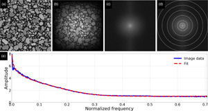
Determining spatial resolution from images is crucial when optimizing focus, determining smallest resolvable object, and assessing size measurement uncertainties. However, no standard algorithm exists to measure resolution from electron microscopy (EM) images, though several have been proposed, where most require user decisions. We present the Spatial Image Resolution Assessment by Fourier analysis (SIRAF) algorithm that uses fast Fourier transform analysis to estimate resolution directly from a single image without user inputs. The method is derived from the underlying assumption that objects display intensity transitions, resembling a step function blurred by a Gaussian point spread function. This hypothesis is tested and verified on simulated EM images with known resolution. To identify potential pitfalls, the algorithm is also tested on simulated images with a variety of settings, and on real SEM images acquired at different magnification and defocus settings. Finally, the versatility of the method is investigated by assessing resolution in images from several microscopy techniques. It is concluded that the algorithm can assess resolution from a large selection of image types, thereby providing a measure of this fundamental image parameter. It may also improve autofocus methods and guide the optimization of magnification settings when balancing spatial resolution and field of view.

Type
Software and Instrumentation
Copyright
Copyright © The Author(s), 2022. Published by Cambridge University Press on behalf of the Microscopy Society of America

Access options

Get access to the full version of this content by using one of the access options below. (Log in options will check for institutional or personal access. Content may require purchase if you do not have access.)

References

Babin, S, Gaevski, M, Joy, D, Machin, M & Martynov, A (2006). Technique to automatically measure electron-beam diameter and astigmatism: BEAMETR. J Vac Sci Technol 24(6), 2956.CrossRefGoogle Scholar
Bradski, G (2000). The OpenCV library. Dr Dobbs J Soft Tools 25, 120125.Google Scholar
Brostrøm, A, Kling, KI, Hougaard, KS & Mølhave, K (2020). Complex aerosol characterization by scanning electron microscopy coupled with energy dispersive X-ray spectroscopy. Sci Rep 10(1). doi:10.1038/s41598-020-65383-5CrossRefGoogle ScholarPubMed
Brostrøm, A, Kling, KI, Koponen, IK, Hougaard, KS, Kandler, K & Mølhave, K (2019). Improving the foundation for particulate matter risk assessment by individual nanoparticle statistics from electron microscopy analysis. Sci Rep 9(1), 8093.CrossRefGoogle ScholarPubMed
Cizmar, P, Vladár, AE, Ming, B & Postek, MT (2008). Simulated SEM images for resolution measurement. Scanning 30(5), 381391.CrossRefGoogle ScholarPubMed
Conti, S, Perico, N, Novelli, R, Carrara, C, Benigni, A & Remuzzi, G (2018). Early and late scanning electron microscopy findings in diabetic kidney disease. Sci Rep 8(1). doi:10.1038/s41598-018-23244-2CrossRefGoogle ScholarPubMed
Crouzier, L, Delvallée, A, Allard, A, Devoille, L, Ducourtieux, S & Feltin, N (2019). Methodology to evaluate the uncertainty associated with nanoparticle dimensional measurements by SEM. Meas Sci Technol 30(8). doi:10.1088/1361-6501/ab1495CrossRefGoogle Scholar
Dalgleish, DG, Spagnuolo, PA & Douglas Goff, H (2004). A possible structure of the casein micelle based on high-resolution field-emission scanning electron microscopy. Int Dairy J 14(12), 10251031.CrossRefGoogle Scholar
Egerton, RF, Li, P & Malac, M (2004). Radiation damage in the TEM and SEM. Micron 35(6), 399409.CrossRefGoogle ScholarPubMed
Erdman, N, Bell, DC & Reichelt, R (2019). Scanning electron microscopy. In Springer Handbooks of Microscopy, Hawkes, PW & Spence, JCH (Eds.), pp. 229318. Cham: Springer.CrossRefGoogle Scholar
Hearle, JWS, Lomas, B & Sparrow, JT (1970). The selection of conditions for examination of specimens in a scanning electron microscope. J Microsc 92(3), 205216.CrossRefGoogle Scholar
Hein, LRO, Campos, KA, Caltabiano, PCRO & Kostov, KG (2013). A brief discussion about image quality and SEM methods for quantitative fractography of polymer composites. Scanning 35(3), 196204.CrossRefGoogle ScholarPubMed
Ishitani, T, Kamiya, C & Sato, M (2005). Influence of random noise on the contrast-to-gradient image resolution in scanning electron microscopy. J Electron Microsc 54(2), 8597.Google ScholarPubMed
Ishitani, T & Sato, M (2002 a). A method for personal expertise-independent evaluation of image resolution in scanning electron microscopy. Scanning 24(4), 191203.CrossRefGoogle ScholarPubMed
Ishitani, T & Sato, M (2002 b). Contrast-to-gradient method for the evaluation of image resolution in scanning electron microscopy. J Electron Microsc 51(6), 369382.CrossRefGoogle ScholarPubMed
Ishitani, T & Sato, M (2004). Contrast-to-gradient method for the evaluation of image resolution taking account of random noise in scanning electron microscopy. J Electron Microsc 53(3), 245255.CrossRefGoogle ScholarPubMed
Ishitani, T & Sato, M (2007). Evaluation of both image resolution and contrast-to-noise ratio in scanning electron microscopy. J Electron Microsc 56(4), 145151.CrossRefGoogle ScholarPubMed
Joy, DC (1991). Contrast in high-resolution scanning electron microscope images. J Microsc 161(2), 343355.CrossRefGoogle Scholar
Joy, DC (1995). A database on electron-solid interactions. Scanning 17(5), 270275.CrossRefGoogle Scholar
Joy, DC (2002). SMART: A program to measure SEM resolution and imaging performance. J Microsc 208(1), 2434.CrossRefGoogle ScholarPubMed
Joy, DC, Ko, Y-U & Hwu, JJ (2000). Metric of resolution and performance for CD-SEMs. In Metrology, Inspection, and Process Control for Microlithography XIV, Sullivan, NT (Ed.), vol. 3998. pp. 108114. Santa Clara, CA, USA: MICROLITHOGRAPHY 2000 (or SPIE).CrossRefGoogle Scholar
Lifshin, E, Kandel, YP & Moore, RL (2014). Improving scanning electron microscope resolution for near planar samples through the use of image restoration. Microscopy and Microanalysis 20(1), 7889.CrossRefGoogle ScholarPubMed
Lifshin, E, Zotta, M, Frey, D, Lifshin, S, Nevins, M & Moskin, J (2017). A software approach to improving SEM resolution, image quality, and productivity. Micros Today 25(3), 1825.CrossRefGoogle Scholar
Lorusso, GF & Joy, DC (2003). Experimental resolution measurement in critical dimension scanning electron microscope metrology. Scanning 25(4), 175180.CrossRefGoogle ScholarPubMed
Maraghechi, S, Hoefnagels, JPM, Peerlings, RHJ & Geers, MGD (2018). Correction of scan line shift artifacts in scanning electron microscopy: An extended digital image correlation framework. Ultramicroscopy 187, 144163.CrossRefGoogle ScholarPubMed
Mizutani, R, Saiga, R, Takekoshi, S, Inomoto, C, Nakamura, N, Itokawa, M, Arai, M, Oshima, K, Takeuchi, A, Uesugi, K, Terada, Y & Suzuki, Y (2016). A method for estimating spatial resolution of real image in the Fourier domain. J Microsc 261(1), 5766.CrossRefGoogle Scholar
Mumpton, FA & Ormsby, WC (1976). Morphology of zeolites in sedimentary rocks by scanning electron microscopy. Clays Clay Miner 24(1), 123.CrossRefGoogle Scholar
Nie, B, Liu, X, Yang, L, Meng, J & Li, X (2015). Pore structure characterization of different rank coals using gas adsorption and scanning electron microscopy. Fuel 158, 908917.CrossRefGoogle Scholar
Nyquist, H (1928). Certain topics in telegraph transmission theory. Trans AIEE 47, 617644.Google Scholar
Ong, KH, Phang, JCH & Thong, JTL (1997). A robust focusing and astigmatism correction method for the scanning electron microscope. Scanning 19(8), 553563.CrossRefGoogle Scholar
Shah, FA, Ruscsák, K & Palmquist, A (2019). 50 years of scanning electron microscopy of bone — A comprehensive overview of the important discoveries made and insights gained into bone material properties in health, disease, and taphonomy. Bone Res 7(1). doi:10.1038/s41413-019-0053-zCrossRefGoogle Scholar
Xing, Q (2016). Information or resolution: Which is required from an SEM to study bulk inorganic materials? Scanning 38(6), 864879.CrossRefGoogle ScholarPubMed
Yesibolati, MN, Mortensen, KI, Sun, H, Brostrøm, A, Tidemand-Lichtenberg, S & Mølhave, K (2020). Unhindered Brownian motion of individual nanoparticles in liquid-phase scanning transmission electron microscopy. Nano Lett 20(10), 71087115.CrossRefGoogle ScholarPubMed
Supplementary material: File

Brostrøm and Mølhave supplementary material

Brostrøm and Mølhave supplementary material 1

Download Brostrøm and Mølhave supplementary material(File)
File 17.5 KB
Supplementary material: File

Brostrøm and Mølhave supplementary material

Brostrøm and Mølhave supplementary material 2

Download Brostrøm and Mølhave supplementary material(File)
File 18.2 KB
Supplementary material: File

Brostrøm and Mølhave supplementary material

Brostrøm and Mølhave supplementary material 3

Download Brostrøm and Mølhave supplementary material(File)
File 6.8 KB
Supplementary material: File

Brostrøm and Mølhave supplementary material

Brostrøm and Mølhave supplementary material 4

Download Brostrøm and Mølhave supplementary material(File)
File 27.9 MB