Hostname: page-component-cd9895bd7-q99xh Total loading time: 0 Render date: 2024-12-23T14:18:36.307Z Has data issue: false hasContentIssue false

Processing APT Spectral Backgrounds for Improved Quantification

Published online by Cambridge University Press:  19 August 2020

Daniel Haley*
Affiliation:
Department of Materials, University of Oxford, Parks Rd, Oxford, OxfordshireOX1 3PH, UK
Andrew J. London
Affiliation:
United Kingdom Atomic Energy Authority, Culham Centre for Fusion Energy, Culham Science Centre, Abingdon, OxonOX14 3DB, UK
Michael P. Moody
Affiliation:
Department of Materials, University of Oxford, Parks Rd, Oxford, OxfordshireOX1 3PH, UK
*
*Author for correspondence: Daniel Haley, E-mail: [email protected]
Get access

Abstract

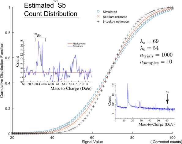
We describe a method to estimate background noise in atom probe tomography (APT) mass spectra and to use this information to enhance both background correction and quantification. Our approach is mathematically general in form for any detector exhibiting Poisson noise with a fixed data acquisition time window, at voltages varying through the experiment. We show that this accurately estimates the background observed in real experiments. The method requires, as a minimum, the z-coordinate and mass-to-charge-state data as input and can be applied retrospectively. Further improvements are obtained with additional information such as acquisition voltage. Using this method allows for improved estimation of variance in the background, and more robust quantification, with quantified count limits at parts-per-million concentrations. To demonstrate applications, we show a simple peak detection implementation, which quantitatively suppresses false positives arising from random noise sources. We additionally quantify the detectability of 121-Sb in a standardized-doped Si microtip as (1.5 × 10⁻⁵, 3.8 × 10⁻⁵) atomic fraction, α = 0.95. This technique is applicable to all modes of APT data acquisition and is highly general in nature, ultimately allowing for improvements in analyzing low ionic count species in datasets.

Type
Software and Instrumentation
Copyright
Copyright © Microscopy Society of America 2020

Access options

Get access to the full version of this content by using one of the access options below. (Log in options will check for institutional or personal access. Content may require purchase if you do not have access.)

References

Alvarez, JL (2007). Poisson-based detection limit and signal confidence intervals for few total counts. J Health Phys 93, 120126.CrossRefGoogle ScholarPubMed
Bityukov, SI, Krasnikov, NV & Taperechkina, VA (2001). Confidence intervals for Poisson distribution parameter. arXiv:hep-ex/0108020v1.Google Scholar
Cerezo, A, Clifton, PH, Galtrey, MJ, Humphreys, CJ, Kelly, TF, Larson, DJ, Lozano-Perez, S, Marquis, EA, Oliver, RA, Sha, G, Thompson, K, Zandbergen, M & Alvis, RL (2007). Atom probe tomography today. Mater Today 10, 3642.CrossRefGoogle Scholar
Cerioli, A & Farcomeni, A (2011). Error rates for multivariate outlier detection. Comput Stat Data Anal 55, 544553.CrossRefGoogle Scholar
Croarkin, C & Tobias, PE (2019). Engineering statistics handbook [Online].Google Scholar
Currie, L (1968). Limits for qualitative detection and quantitative determination: Application to radiochemistry. Anal Chem 3, 586593.CrossRefGoogle Scholar
Dong, Y, Motta, A & Marquis, E (2013). Atom probe tomography study of alloying element distributions in Zr alloys and their oxides. J Nucl Mater 442, 270281.CrossRefGoogle Scholar
Giddings, AD, Koelling, S, Shimizu, Y, Estivill, R, Inoue, K, Vandervorst, W & Yeoh, WK (2018). Industrial application of atom probe tomography to semiconductor devices. Scr Mater 148, 8290.CrossRefGoogle Scholar
Haley, D, Choi, P & Raabe, D (2015). Guided mass spectrum labelling in atom probe tomography. Ultramicroscopy 159, 338345.CrossRefGoogle ScholarPubMed
Haley, D, Merzlikin, S, Choi, P & Raabe, D (2014). Atom probe tomography observation of hydrogen in high-Mn steel and silver charged via an electrolytic route. Int J Hydrogen Energy 39, 1222112229.CrossRefGoogle Scholar
Heck, PR, Stadermann, FJ, Isheim, D, Auciello, O, Daulton, TL, Davis, AM, Elam, JW, Floss, C, Hiller, J, Larson, DJ, Lewis, J, Mane, A, Pellin, MJ, Savina, MR, Seidman, DN & Stephan, T (2014). Atom-probe analyses of nanodiamonds from Allende. Meteorit Planet Sci 49, 453467.CrossRefGoogle Scholar
Hudson, D, Smith, G & Gault, B (2011). Optimisation of mass ranging for atom probe microanalysis and application to the corrosion processes in Zr alloys. Ultramicroscopy 111, 480486.CrossRefGoogle ScholarPubMed
Larson, DJ, Prosa, T, Ulfig, R, Geiser, B & Kelly, T (2014). Local Electrode Atom Probe Tomography. New York: Springer.Google Scholar
London, AJ, Haley, D & Moody, MP (2017). Single-ion deconvolution of mass peak overlaps for atom probe microscopy. Microsc Microanal 23, 300306.CrossRefGoogle ScholarPubMed
Meier, M (2019). Developing laser-pulsed atom probe tomography techniques for 3D imaging of hydrogen in steel. Master's Thesis. Friedrich-Alexander Universität Erlangen-Nürnberg. arXiv:submit/3177611.Google Scholar
Oltman, E, Ulfig, RM & Larson, DJ (2009). Background removal methods applied to atom probe data. Microsc Microanal 15, 256257.CrossRefGoogle Scholar
Piazolo, S, La Fontaine, A, Trimby, P, Harley, S, Yang, L, Armstrong, R & Cairney, JM (2016). Deformation-induced trace element redistribution in zircon revealed using atom probe tomography. Nat Commun 7, 10490.CrossRefGoogle ScholarPubMed
Saxey, DW, Moser, DE, Piazolo, S, Reddy, S & Valley, J (2018). Atomic worlds: Current state and future of atomprobe microscopy in geoscience. Scr Mater 148, 115121.CrossRefGoogle Scholar
Tu, Y, Han, B, Shimizu, Y, Inoue, K, Fukui, Y, Yano, M, Tanii, T, Shinada, T & Nagai, Y (2017). Atom probe tomographic assessment of the distribution of germanium atoms implanted in a silicon matrix through nano-apertures. Nanotechnology 28, 385301.CrossRefGoogle Scholar
Zech, G (1989). Upper limits in experiments with background or measurement errors. Nucl Instrum Meth Phys Res A 277, 608610.CrossRefGoogle Scholar