Hostname: page-component-586b7cd67f-t8hqh Total loading time: 0 Render date: 2024-11-22T18:32:02.509Z Has data issue: false hasContentIssue false

Microanalysis of the Intestinal Bulb of Grass Carp (Ctenopharyngodon Idella): Histological, Histochemical, Immunohistochemical, and Scanning Electron Microscopical Studies

Published online by Cambridge University Press:  06 October 2021

Doaa M. Mokhtar*
Affiliation:
Department of Anatomy and Histology, Faculty of Veterinary Medicine, Assuit University, 71526Assiut, Egypt
Enas A. Abd-Elhafez
Affiliation:
Department of Anatomy and Histology, Faculty of Veterinary Medicine, Assuit University, 71526Assiut, Egypt
Ahmed H. S. Hassan
Affiliation:
Department of Anatomy and Histology, Faculty of Veterinary Medicine, Assuit University, 71526Assiut, Egypt
*
*Corresponding author: Doaa M. Mokhtar, E-mail: [email protected]; [email protected]
Get access

Abstract

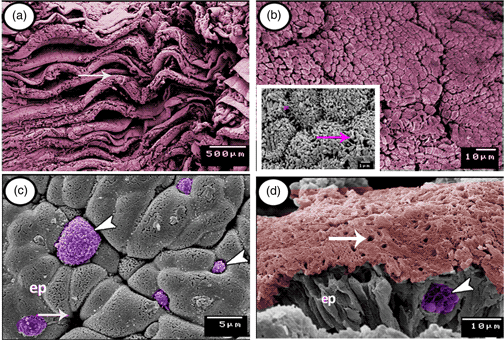
Cyprinid fishes have one of the simplest types of gastrointestinal tract among vertebrates. Those fish species do not possess a true stomach that is replaced by a simple dilatation at the anterior part of the intestine called the intestinal bulb. Twenty adult specimens of grass carp were used in the present study to identify the cellular components as well as the immunohistochemical and surface architectural characteristics of the intestinal bulb. The mucosa of the intestinal bulb shows numerous, deep longitudinal folds arranged in zigzagging-like patterns. The epithelium is composed mainly of absorptive columnar cells covered by microvilli and mucous goblet cells. Spindle-shaped enteroendocrine cells and some migratory immune cells such as intraepithelial lymphocytes and rodlet cells could be identified between the absorptive cells. The epithelium also contains many secretory granules and large numbers of vacuoles containing digestive enzymes mostly in the basal part. The immunohistochemistry revealed that CD20-positive B-lymphocytes are immunolocalized mainly in the connective tissue core lamina propria of the mucosal folds. However, CD3-immunopositive T-lymphocytes are highly concentrated in the lamina propria. In addition, intraepithelial T-lymphocytes expressed immunopositivity to CD3. The current study presented many types of immune cells and suggests their essential immunological role for the intestinal blub.

Type
Micrographia
Copyright
Copyright © The Author(s), 2021. Published by Cambridge University Press on behalf of the Microscopy Society of America

Access options

Get access to the full version of this content by using one of the access options below. (Log in options will check for institutional or personal access. Content may require purchase if you do not have access.)

References

Abdulhadi, HA (2005). Some comparative histological studies on alimentary tract of tilapia fish (Tilapia spilurus) and sea bream (Mylio cuvieri). Egyptian J Aquatic Res 31, 387397.Google Scholar
Albrecht, MP, Ferreira, MFN & Caramaschi, EP (2001). Anatomical features and histology of the digestive tract of two related neotropical omnivorous fishes (Characiformes; Anostomidae). J Fish Biology 58, 419430.CrossRefGoogle Scholar
Bancroft, JD & Steven, A (1996). Theory and Practice of Histological Techniques, 4th ed. New York, Edinburgh, London, Madrid, Melbourne, San Francisco, Tokyo: Churchill Livingstone.Google Scholar
Bernard, D, Six, A, Rigottier-Gois, L, Messiaen, S, Chilmonczyk, S, Quillet, E, Boudinot, P & Benmansour, A (2006). Phenotypic and functional similarity of gut intraepithelial and systemic T cells in a teleost fish. J Immunol 176, 39423949.CrossRefGoogle Scholar
Caceci, T (1984). Scanning electron microscopy of goldfish, Carassius auratus, intestinal mucosa. J Fish Biol 25, 112.CrossRefGoogle Scholar
Dezfuli, BS, Capuano, S & Manera, M (1998). A description of rodlet cells from the alimentary canal of Anguilla anguilla and their relationship with parasitic helminths. J Fish Biol 53, 10841095.Google Scholar
Fiertak, A & Kilarski, WM (2002). Glycoconjugates of the intestinal goblet cells of four cyprinids. J Cell Mol Life Sci 59, 17241733.CrossRefGoogle ScholarPubMed
Fischer, U, Koppang, EO & Nakanishi, T (2013). Teleost T and NK cell immunity. Fish Shellfish Immunol 35, 197206.CrossRefGoogle Scholar
Fischer, U, Utke, K, Somamoto, T, Köllner, B, Ototake, M & Nakanishi, T (2006). Cytotoxic activities of fish leucocytes. Fish Shellfish Immunol 20, 209226.CrossRefGoogle ScholarPubMed
Fugi, R, Agostinho, AA & Hahn, NS (2001). Trophic morphology of five benthic-feeding fish species of a tropical floodplain. Rev Bras Biol 61, 112120.CrossRefGoogle ScholarPubMed
Gross, CA, Reddy, CK & Dazzo, FB (2010). CMEIAS color segmentation: An improved computing technology to process color images for quantitative microbial ecology studies at single-cell resolution. Microb Ecol 59, 400414.CrossRefGoogle ScholarPubMed
Hernandez, DR, Gianeselli, PM & Domitrovic, HA (2009). Morphology, histology and histochemistry of the digestive system of South American Catfish (Rhamdia quelen). J Int Morphol 27, 105111.CrossRefGoogle Scholar
Hsu, SM, Raine, L & Fanger, H (1981). Use of avidin-biotin-peroxidase complex (ABC) in immunoperoxidase techniques: A comparison between ABC and unlabeled antibody (PAP) procedures. J Histochem Cytochem 29, 577580.CrossRefGoogle ScholarPubMed
Kaattari, SL (1992). Fish B lymphocytes: Defining their form and function. Annual Review of Fish Diseases 2, 161180.CrossRefGoogle Scholar
Karnovsky, MJ (1965). A formaldehyde glutaraldehyde fixative of high osmolality for use in electron microscopy. J Cell Biol 27, 137138.Google Scholar
Manjakasy, JM, Day, RD, Kemp, A & Tibbetts, IR (2009). Functional morphology of digestion in the stomachless, piscivorous needlefishes Tylosurus gavialoides and Strongylura leiura ferox (Teleostei: Beloniformes). J Morphol 270, 11551165.CrossRefGoogle Scholar
Mir, IH & Channa, A (2010). A scanning electron microscopic examination of the intestinal Schizothorax curvifrons heckel. J Fish Aquat Sci 5, 386393.Google Scholar
Mishra, KP & Ahmed, MF (1991). Histology and histochemistry of the stomach and intestinal bulb of three hillstream losches. Func Dev Morphol 1, 1114.Google ScholarPubMed
Mokhtar, DM (2017). Fish Histology from Cells to Organs, 1st ed. Canada: Apple Academic Press.CrossRefGoogle Scholar
Mokhtar, DM (2019). Characterization of the fish ovarian stroma during the spawning season: Cytochemical, immunohistochemical and ultrastructural studies. Fish Shellfish Immunol 94, 566579.CrossRefGoogle ScholarPubMed
Mokhtar, DM (2021). Fish Histology from Cells to Organs, 2nd ed. Canada: Apple Academic Press.CrossRefGoogle Scholar
Mokhtar, DM, Abd-Elhafez, EA & Hassan, AHS (2017). Histology, histochemistery and surface architecture of the rectum of grass carp (Ctenopharyngodon idella). J Advanced Micros Res 12, 15.CrossRefGoogle Scholar
Morrison, CM & Wright, JR (1999). A study of the histology of the digestive tract of Nile tilapia. J Fish Biol 54, 597606.CrossRefGoogle Scholar
Pan, QS, Fang, ZP & Zhao, YX (2000). Immunocytochemical identification and localization of APUD cells in the gut of seven stomachless teleost fishes. J World Gastroenterol 6, 96101.CrossRefGoogle ScholarPubMed
Pileri, SA, Roncador, G, Ceccarelli, C, Piccioli, M, Briskomatis, A, Sabattini, E, Ascani, S, Santini, D, Piccaluga, PP, Leone, O, Damiani, S, Ercolessi, C, Sandri, F, Pieri, F, Leoncini, L & Falini, B (1997). Antigen retrieval techniques in immunohistochemistry: Comparison of different methods. J Pathol 183(1), 116123.3.0.CO;2-2>CrossRefGoogle ScholarPubMed
Rombout, JH, Yang, G & Kiron, V (2014). Adaptive immune responses at mucosal surfaces of teleost fish. Fish Shellfish Immunol 40(2), 634643.CrossRefGoogle ScholarPubMed
Stroband, HWJ & Debets, FMH (1978). The ultrastructure and renewal of the intestinal epithelium of the juvenile grass carp (Ctenopharyngodon idella, Val.). J Cell Tissue Res 187, 181200.Google Scholar
Yamamoto, T (1966). An electron microscope study of the columnar epithelial cell in the intestine of the fresh water teleosts: Goldfish (Carassius auratus) and rainbow trout (Salmo irideus). Z Zellforsch Mikrosk Anat 72, 6687.CrossRefGoogle Scholar
Zapata, A, Diez, B, Cejalvo, T, Gutiérrez-de Frías, C & Cortés, A (2006). Ontogeny of the immune system of fish. Fish Shellfish Immunol 20, 126136.CrossRefGoogle ScholarPubMed