Hostname: page-component-586b7cd67f-g8jcs Total loading time: 0 Render date: 2024-11-25T09:22:15.824Z Has data issue: false hasContentIssue false

ESEM Methodology for the Study of Ice Samples at Environmentally Relevant Subzero Temperatures: “Subzero ESEM”

Published online by Cambridge University Press:  23 December 2021
Kamila Závacká*
Affiliation:
Environmental Electron Microscopy Group, Institute of Scientific Instruments of the Czech Academy of Sciences, Královopolská 147, 61264Brno, Czech Republic
Vilém Neděla
Affiliation:
Environmental Electron Microscopy Group, Institute of Scientific Instruments of the Czech Academy of Sciences, Královopolská 147, 61264Brno, Czech Republic
Eva Tihlaříková
Affiliation:
Environmental Electron Microscopy Group, Institute of Scientific Instruments of the Czech Academy of Sciences, Královopolská 147, 61264Brno, Czech Republic
Pavla Šabacká
Affiliation:
Environmental Electron Microscopy Group, Institute of Scientific Instruments of the Czech Academy of Sciences, Královopolská 147, 61264Brno, Czech Republic
Jiří Maxa
Affiliation:
Environmental Electron Microscopy Group, Institute of Scientific Instruments of the Czech Academy of Sciences, Královopolská 147, 61264Brno, Czech Republic
Dominik Heger
Affiliation:
Department of Chemistry, Faculty of Science, Masaryk University, Kamenice 5, 62500Brno, Czech Republic
*
*Corresponding author: Kamila Závacká, E-mail: [email protected]
Get access

Abstract

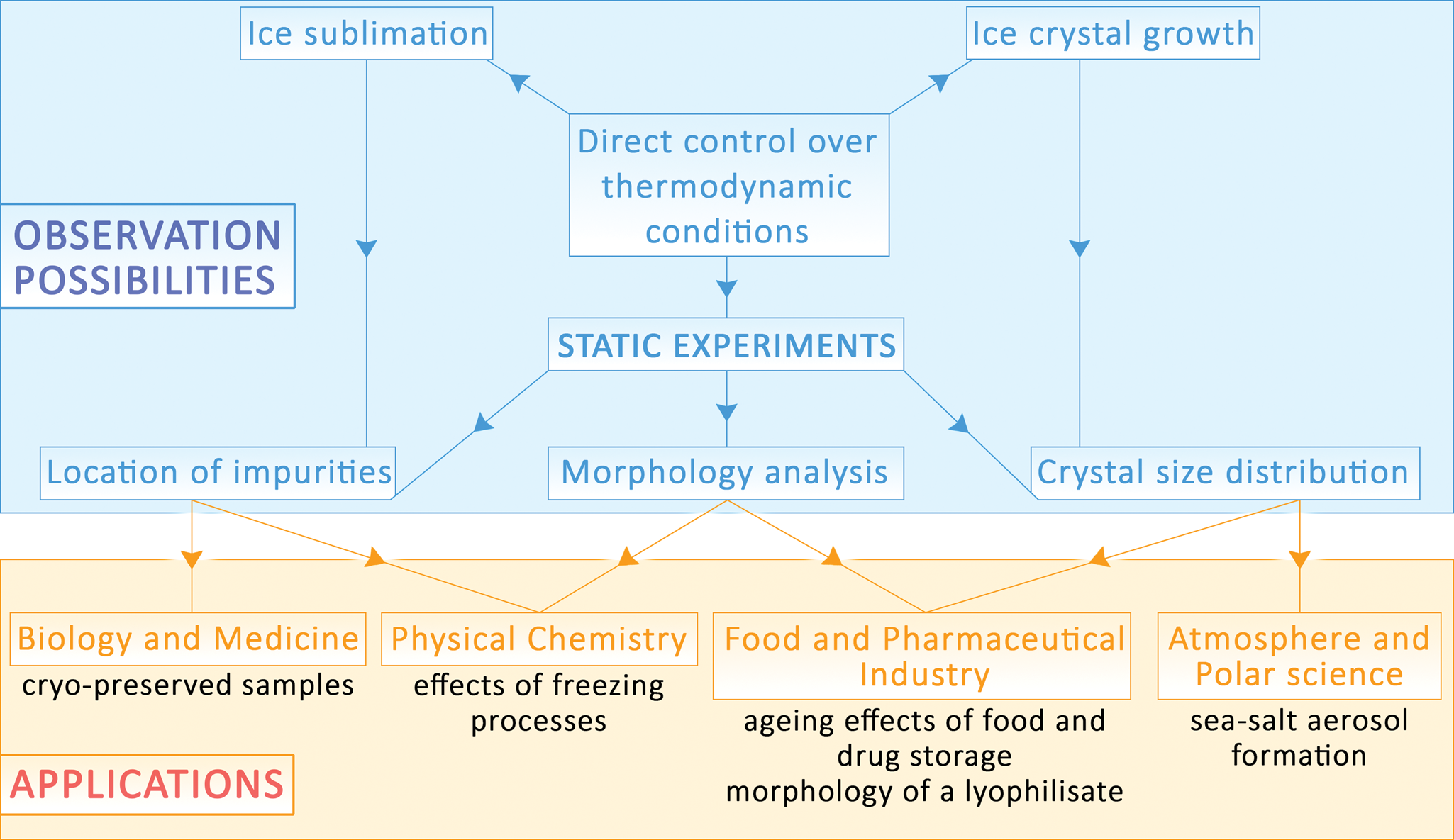
Frozen aqueous solutions are an important subject of study in numerous scientific branches including the pharmaceutical and food industry, atmospheric chemistry, biology, and medicine. Here, we present an advanced environmental scanning electron microscope methodology for research of ice samples at environmentally relevant subzero temperatures, thus under conditions in which it is extremely challenging to maintain the thermodynamic equilibrium of the specimen. The methodology opens possibilities to observe intact ice samples at close to natural conditions. Based on the results of ANSYS software simulations of the surface temperature of a frozen sample, and knowledge of the partial pressure of water vapor in the gas mixture near the sample, we monitored static ice samples over several minutes. We also discuss possible artifacts that can arise from unwanted surface ice formation on, or ice sublimation from, the sample, as a consequence of shifting conditions away from thermodynamic equilibrium in the specimen chamber. To demonstrate the applicability of the methodology, we characterized how the true morphology of ice spheres containing salt changed upon aging and the morphology of ice spheres containing bovine serum albumin. After combining static observations with the dynamic process of ice sublimation from the sample, we can attain images with nanometer resolution.

Type
Software and Instrumentation
Copyright
Copyright © The Author(s), 2021. Published by Cambridge University Press on behalf of the Microscopy Society of America

Access options

Get access to the full version of this content by using one of the access options below. (Log in options will check for institutional or personal access. Content may require purchase if you do not have access.)

References

Adams, EE, Miller, DA & Brown, RL (2001). Grain boundary ridge on sintered bonds between ice crystals. J Appl Phys 90, 57825785.CrossRefGoogle Scholar
Agostinelli, S, Allison, J, Amako, K, Apostolakis, J, Araujo, H, Arce, P, Asai, M, Axen, D, Banerjee, S, Barrand, G, Behner, F, Bellagamba, L, Boudreau, J, Broglia, L, Brunengo, A, Burkhardt, H, Chauvie, S, Chuma, J, Chytracek, R, Cooperman, G, Cosmo, G, Degtyarenko, P, Dell'Acqua, A, Depaola, G, Dietrich, D, Enami, R, Feliciello, A, Ferguson, C, Fesefeldt, H, Folger, G, Foppiano, F, Forti, A, Garelli, S, Giani, S, Giannitrapani, R, Gibin, D, Gomez Cadenas, JJ, Gonzalez, I, Gracia Abril, G, Greeniaus, G, Greiner, W, Grichine, V, Grossheim, A, Guatelli, S, Gumplinger, P, Hamatsu, R, Hashimoto, K, Hasui, H, Heikkinen, A, Howard, A, Ivanchenko, V, Johnson, A, Jones, FW, Kallenbach, J, Kanaya, N, Kawabata, M, Kawabata, Y, Kawaguti, M, Kelner, S, Kent, P, Kimura, A, Kodama, T, Kokoulin, R, Kossov, M, Kurashige, H, Lamanna, E, Lampen, T, Lara, V, Lefebure, V, Lei, F, Liendl, M, Lockman, W, Longo, F, Magni, S, Maire, M, Medernach, E, Minamimoto, K, Mora de Freitas, P, Morita, Y, Murakami, K, Nagamatu, M, Nartallo, R, Nieminen, P, Nishimura, T, Ohtsubo, K, Okamura, M, O'Neale, S, Oohata, Y, Paech, K, Perl, J, Pfeiffer, A, Pia, MG, Ranjard, F, Rybin, A, Sadilov, S, di Salvo, E, Santin, G, Sasaki, T, Savvas, N, Sawada, Y, Scherer, S, Sei, S, Sirotenko, V, Smith, D, Starkov, N, Stoecker, H, Sulkimo, J, Takahata, M, Tanaka, S, Tcherniaev, E, Safai Tehrani, E, Tropeano, M, Truscott, P, Uno, H, Urban, L, Urban, P, Verderi, M, Walkden, A, Wander, W, Weber, H, Wellisch, JP, Wenaus, T, Williams, DC, Wright, D, Yamada, T, Yoshida, H & Zschiesche, D (2003). GEANT4 - A simulation toolkit. Nucl Instrum Methods Phys Res A 506, 250303.CrossRefGoogle Scholar
Aider, M & de Halleux, D (2009). Cryoconcentration technology in the bio-food industry: Principles and applications. Lebensm Wiss Technol 42, 679685.CrossRefGoogle Scholar
Authelin, JR, Rodrigues, MA, Tchessalov, S, Singh, SK, McCoy, T, Wang, S & Shalaev, E (2020). Freezing of biologicals revisited: Scale, stability, excipients, and degradation stresses. J Pharm Sci 109, 4461.10.1016/j.xphs.2019.10.062CrossRefGoogle ScholarPubMed
Barnes, PRF, Wolff, EW, Mallard, DC & Mader, HM (2003). SEM studies of the morphology and chemistry of polar ice. Microsc Res Tech 62, 6269.CrossRefGoogle ScholarPubMed
Bartels-Rausch, T, Jacobi, HW, Kahan, TF, Thomas, JL, Thomson, ES, Abbatt, JPD, Ammann, M, Blackford, JR, Bluhm, H, Boxe, C, Domine, F, Frey, MM, Gladich, I, Guzmán, MI, Heger, D, Huthwelker, T, Klán, P, Kuhs, WF, Kuo, MH, Maus, S, Moussa, SG, McNeill, VF, Newberg, JT, Pettersson, JBC, Roeselová, M & Sodeau, JR (2014). A review of air–ice chemical and physical interactions (AICI): Liquids, quasi-liquids, and solids in snow. Atmos Chem Phys 14, 15871633.CrossRefGoogle Scholar
Beckett, A & Read, ND (1986). Low-temperature scanning electron microscopy. In Ultrastructure Techniques for Microorganisms, Aldrich, HC & Todd, W (Eds.), pp. 4585. New York: Plenum Press.CrossRefGoogle Scholar
Blackford, JR (2007). Sintering and microstructure of ice: A review. J Phys D Appl Phys 40, R355R385.CrossRefGoogle Scholar
Blackford, JR, Jeffree, CE, Noake, DFJ & Marmo, BA (2007). Microstructural evolution in sintered ice particles containing NaCl observed by low-temperature scanning electron microscope. Proc Inst Mech Eng L 221, 151156.Google Scholar
Bomben, JL & King, CJ (1982). Heat and mass transport in the freezing of apple tissue. Int J Food Sci Technol 17, 615632.10.1111/j.1365-2621.1982.tb00221.xCrossRefGoogle Scholar
Cameron, RE & Donald, AM (1994). Minimizing sample evaporation in the environmental scanning electron microscope. J Microsc 173, 227237.CrossRefGoogle Scholar
Chen, X, Shu, J & Chen, Q (2017). Abnormal gas-liquid-solid phase transition behaviour of water observed with in situ environmental SEM. Sci Rep 7, 46680.10.1038/srep46680CrossRefGoogle ScholarPubMed
Craig, S & Beaton, CD (1996). A simple cryo-SEM method for delicate plant tissues. J Microsc 182, 102105.CrossRefGoogle Scholar
Cullen, D & Baker, I (2001). Observation of impurities in ice. Microsc Res Tech 55, 198207.CrossRefGoogle ScholarPubMed
Danilatos, GD (1981). Design and construction of an atmospheric or environmental SEM (part 1). Scanning 4, 920.CrossRefGoogle Scholar
Danilatos, GD (2013). Electron scattering cross-section measurements in ESEM. Micron 45, 116.CrossRefGoogle ScholarPubMed
Donald, AM (2003). The use of environmental scanning electron microscopy for imaging wet and insulating materials. Nat Mater 2, 511516.CrossRefGoogle ScholarPubMed
Du, J, Pushkarova, RA & Smart, RSC (2009). A cryo-SEM study of aggregate and floc structure changes during clay settling and raking processes. Int J Miner Process 93, 6672.CrossRefGoogle Scholar
Fletcher, AL, Keller, TH, Thiel, BL, Eddy, AE & Donald, AM (1998). ‘Cool ESEM’ – imaging ice-containing systems at freezer temperatures. Microsc Microanal 4 (Suppl 2), 284285.CrossRefGoogle Scholar
Fourie, JT (1982). Gold in electron microscopy. Gold Bull 15, 26.CrossRefGoogle Scholar
Grothe, H, Baloh, P, Whitmore, K & Waller, D (2009). Environmental scanning electron microscopy (ESEM) of atmospheric ices. Geophys Res Abstr 11, 5905.Google Scholar
Heldner, M (2014). Pharmaceutical freeze-drying systems. In Vacuum Technology in the Chemical Industry, Jorisch, W (Ed.), pp. 259279. Germany: Wiley-VCH Verlag.Google Scholar
Hullar, T & Anastasio, C (2016). Direct visualization of solute locations in laboratory ice samples. Cryosphere 10, 20572068.10.5194/tc-10-2057-2016CrossRefGoogle Scholar
Imrichova, K, Veselý, L, Gasser, TM, Loerting, T, Neděla, V & Heger, D (2019). Vitrification and increase of basicity in between ice Ih crystals in rapidly frozen dilute NaCl aqueous solutions. J Chem Phys 151, 014503.CrossRefGoogle ScholarPubMed
Issman, L & Talmon, Y (2012). Cryo-SEM specimen preparation under controlled temperature and concentration conditions. J Microsc 246, 6069.CrossRefGoogle ScholarPubMed
John Morris, G & Acton, E (2013). Controlled ice nucleation in cryopreservation: A review. Cryobiology 66, 8592.CrossRefGoogle Scholar
Jones, C. G. (2012). Scanning electron microscopy: Preparation and imaging for SEM. In Forensic Microscopy for Skeletal Tissues: Methods and Protocolsvol, vol. 915, Bell, LS (Ed.), pp. 120. Totowa, NJ: Humana Press.CrossRefGoogle Scholar
Keyser, LF & Leu, M-T (1993). Morphology of nitric acid and water ice films. Microsc Res Tech 25, 434438.CrossRefGoogle ScholarPubMed
Kiani, H & Sun, DW (2011). Water crystallization and its importance to freezing of foods: A review. Trends Food Sci Technol 22, 407426.CrossRefGoogle Scholar
Krausko, J, Runštuk, J, Neděla, V, Klán, P & Heger, D (2014). Observation of a brine layer on an ice surface with an environmental scanning electron microscope at higher pressures and temperatures. Langmuir 30, 54415447.CrossRefGoogle Scholar
Li, B & Sun, D-W (2002). Novel methods for rapid freezing and thawing of foods – a review. J Food Eng 54, 175182.CrossRefGoogle Scholar
Lo, CW, Sahoo, V & Lu, MC (2017). Control of ice formation. ACS Nano 11, 26652674.CrossRefGoogle ScholarPubMed
Mccarthy, C, Blackford, JR & Jeffree, CE (2013). Low-temperature-SEM study of dihedral angles in the ice-I/sulfuric acid partially molten system. J Microsc 249, 150157.CrossRefGoogle ScholarPubMed
Michaloudi, E, Papakostas, S, Stamou, G, Neděla, V, Tihlaříková, E, Zhang, W & Declerck, SAJ (2018). Reverse taxonomy applied to the brachionus calyciflorus cryptic species complex: Morphometric analysis confirms species delimitations revealed by molecular phylogenetic analysis and allows the (re) description of four species. PLoS ONE 13, 125.CrossRefGoogle Scholar
Murphy, DM & Koop, T (2005). Review of the vapour pressures of ice and supercooled water for atmospheric applications. Q J R Metereol Soc 131, 15391565.CrossRefGoogle Scholar
Nair, M, Husmann, A, Cameron, RE & Best, SM (2018). In situ ESEM imaging of the vapor-pressure-dependent sublimation-induced morphology of ice. Phys Rev Mater 2, 040401.CrossRefGoogle Scholar
Neděla, V (2010). Controlled dehydration of a biological sample using an alternative form of environmental SEM. J Microsc 237, 711.CrossRefGoogle ScholarPubMed
Neděla, V, Tihlaříková, E & Hřib, J (2015). The low-temperature method for study of coniferous tissues in the environmental scanning electron microscope. Microsc Res Tech 78, 1321.CrossRefGoogle ScholarPubMed
Neděla, V, Tihlaříková, E, Maxa, J, Imrichová, K, Bučko, M & Gemeiner, P (2020). Simulation-based optimisation of thermodynamic conditions in the ESEM for dynamical in-situ study of spherical polyelectrolyte complex particles in their native state. Ultramicroscopy 211, 112954.10.1016/j.ultramic.2020.112954CrossRefGoogle ScholarPubMed
Neděla, V, Tihlaříková, E, Runštuk, J & Hudec, J (2018). High-efficiency detector of secondary and backscattered electrons for low-dose imaging in the ESEM. Ultramicroscopy 184, 111.CrossRefGoogle ScholarPubMed
Pedersen, C, Mihranyan, A & Strømme, M (2011). Surface transition on ice induced by the formation of a grain boundary. PLoS ONE 6, e24373.CrossRefGoogle ScholarPubMed
Petrenko, VF & Whitworth, RW (1999). Physics of Ice. Oxford: Oxford University Press.Google Scholar
Reimer, L (1998). Scanning Electron Microscopy: Physics of Image Formation and Microanalysis. Berlin, Heidelberg, New York, Tokyo: Springer.CrossRefGoogle Scholar
Schenk, O, Urai, JL & Piazolo, S (2006). Structure of grain boundaries in wet, synthetic polycrystalline, statically recrystallizing halite – Evidence from cryo-SEM observations. Geofluids 6, 93104.CrossRefGoogle Scholar
Shalaev, EY & Franks, F (1996). Changes in the physical state of model mixtures during freezing and drying: Impact on product quality. Cryobiology 33, 1426.CrossRefGoogle Scholar
Stelate, A, Tihlaříková, E, Schwarzerová, K, Neděla, V & Petrášek, J (2021). Correlative light-environmental scanning electron microscopy of plasma membrane efflux carriers of plant hormone auxin. Biomolecules 11, 113.CrossRefGoogle ScholarPubMed
Stokes, DJ, Mugnier, JY & Clarke, CJ (2004). Static and dynamic experiments in cryo-electron microscopy: Comparative observations using high-vacuum, low-voltage and low-vacuum SEM. J Microsc 213, 198204.CrossRefGoogle ScholarPubMed
Thiel, BL, Toth, M, Schroemges, RPM, Scholtz, JJ, Van Veen, G & Knowles, WR (2006). Two-stage gas amplifier for ultrahigh resolution low vacuum scanning electron microscopy. Rev Sci Instrum 77, 033705.CrossRefGoogle Scholar
Tihlaříková, E, Neděla, V & Đorđević, B (2019). In-situ preparation of plant samples in ESEM for energy dispersive x-ray microanalysis and repetitive observation in SEM and ESEM. Sci Rep 9, 2300.CrossRefGoogle ScholarPubMed
Varanasi, KK, Deng, T, Smith, JD, Hsu, M & Bhate, N (2010). Frost formation and ice adhesion on superhydrophobic surfaces. Appl Phys Lett 97, 234102.CrossRefGoogle Scholar
Vetráková, Ľ, Neděla, V, Runštuk, J & Heger, D (2019). The morphology of ice and liquid brine in the environmental scanning electron microscope: A study of the freezing methods. The Cryosphere 13, 23852405.CrossRefGoogle Scholar
Vetráková, Ľ, Neděla, V, Runštuk, J, Tihlaříková, E, Heger, D & Shalaev, E (2020). Dynamical in-situ observation of the lyophilization and vacuum-drying processes of a model biopharmaceutical system by an environmental scanning electron microscope. Int J Pharm 585, 119448.CrossRefGoogle Scholar
Vlašínová, H, Neděla, V, Đorđević, B & Havel, L (2017). Bottlenecks in bog pine multiplication by somatic embryogenesis and their visualization with the environmental scanning electron microscope. Protoplasma 254, 14871497.CrossRefGoogle ScholarPubMed
Waller, D, Stokes, DJ & Donald, AM (2008). Improvements to a cryosystem to observe ice nucleating in a variable pressure scanning electron microscope. Rev Sci Instrum 79, 103709.CrossRefGoogle Scholar
Walther, P, Wehrli, E, Hermann, R & Mueller, M (1995). Double-layer coating for high-resolution, low-temperature scanning electron microscopy. J Microsc 179, 229237.CrossRefGoogle Scholar
Wang, B, Knopf, DA, China, S, Arey, BW, Harder, TH, Gilles, MK & Laskin, A (2016). Direct observation of ice nucleation events on individual atmospheric particles. Phys Chem Chem Phys 18, 2972129731.CrossRefGoogle ScholarPubMed
Wang, J, Memon, H, Liu, J, Yang, G, Xu, F, Hussain, T, Scotchford, C & Hou, X (2019). Effect of surface adsorption on icing behaviour of metallic coating. Surf Coat Technol 380, 125068.CrossRefGoogle Scholar
Weikusat, I, de Winter, DAM, Pennock, GM, Hayles, M, Schneijdenberg, CTWM & Drury, MR (2011). Cryogenic EBSD on ice: Preserving a stable surface in a low pressure SEM. J Microsc 242, 295310.CrossRefGoogle Scholar
Wergin, WP, Rango, A & Erbe, EF (1995). Observations of snow crystals using low-temperature scanning electron microscopy. Scanning 17, 4150.CrossRefGoogle Scholar
Wexler, A (1977). Vapor pressure formulation for Ice. J Res Natl Bur Stand A Phys Chem 81A, 520.CrossRefGoogle Scholar
Wexler, A & Greenspan, L (1976). Vapor pressure equation for water in the range 0 to 100 °C. A revision. J Res Natl Bur Stand A Phys Chem 80A, 775785.CrossRefGoogle Scholar
Yang, X, Neděla, V, Runštuk, J, Ondrušková, G, Krausko, J, Vetráková, L & Heger, D (2017). Evaporating brine from frost flowers with electron microscopy and implications for atmospheric chemistry and sea-salt aerosol formation. Atmos Chem Phys 17, 62916303.CrossRefGoogle Scholar
Zimmermann, F, Ebert, M, Worringen, A, Schütz, L & Weinbruch, S (2007). Environmental scanning electron microscopy (ESEM) as a new technique to determine the ice nucleation capability of individual atmospheric aerosol particles. Atmos Environ 41, 82198227.CrossRefGoogle Scholar
Zimmermann, F, Weinbruch, S, Schütz, L, Hofmann, H, Ebert, M, Kandler, K & Worringen, A (2008). Ice nucleation properties of the most abundant mineral dust phases. J Geophys Res Atmos 113, D23204.10.1029/2008JD010655CrossRefGoogle Scholar
Supplementary material: File

Závacká et al. supplementary material

Závacká et al. supplementary material 1

Download Závacká et al. supplementary material(File)
File 965.1 KB

Závacká et al. supplementary material

Závacká et al. supplementary material 2

Download Závacká et al. supplementary material(Video)
Video 6.5 MB

Závacká et al. supplementary material

Závacká et al. supplementary material 3

Download Závacká et al. supplementary material(Video)
Video 4.1 MB