Hostname: page-component-586b7cd67f-rcrh6 Total loading time: 0 Render date: 2024-11-26T07:16:26.209Z Has data issue: false hasContentIssue false

Development of a Practicable Digital Pulse Read-Out for Dark-Field STEM

Published online by Cambridge University Press:  18 December 2020

Tiarnan Mullarkey*
Affiliation:
School of Physics, Trinity College Dublin, Dublin 2, Ireland Centre for Doctoral Training in the Advanced Characterisation of Materials, AMBER Centre, Dublin 2, Ireland
Clive Downing
Affiliation:
Advanced Microscopy Laboratory, Centre for Research on Adaptive Nanostructures and Nanodevices (CRANN), Dublin 2, Ireland
Lewys Jones
Affiliation:
School of Physics, Trinity College Dublin, Dublin 2, Ireland Advanced Microscopy Laboratory, Centre for Research on Adaptive Nanostructures and Nanodevices (CRANN), Dublin 2, Ireland
*
*Author for correspondence: Tiarnan Mullarkey, E-mail: [email protected]
Get access

Abstract

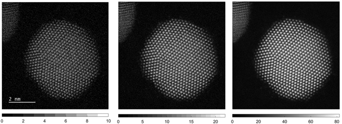
When characterizing beam-sensitive materials in the scanning transmission electron microscope (STEM), low-dose techniques are essential for the reliable observation of samples in their true state. A simple route to minimize both the total electron-dose and the dose-rate is to reduce the electron beam-current and/or raster the probe at higher speeds. At the limit of these settings, and with current detectors, the resulting images suffer from unacceptable artifacts, including signal-streaking, detector-afterglow, and poor signal-to-noise ratios (SNRs). In this article, we present an alternative approach to capture dark-field STEM images by pulse-counting individual electrons as they are scattered to the annular dark-field (ADF) detector. Digital images formed in this way are immune from analog artifacts of streaking or afterglow and allow clean, high-SNR images to be obtained even at low beam-currents. We present results from both a ThermoFisher FEI Titan G2 operated at 300 kV and a Nion UltraSTEM200 operated at 200 kV, and compare the images to conventional analog recordings. ADF data are compared with analog counterparts for each instrument, a digital detector-response scan is performed on the Titan, and the overall rastering efficiency is evaluated for various scanning parameters.

Type
Software and Instrumentation
Copyright
Copyright © The Author(s), 2020. Published by Cambridge University Press

Access options

Get access to the full version of this content by using one of the access options below. (Log in options will check for institutional or personal access. Content may require purchase if you do not have access.)

References

Béché, A, Goris, B, Freitag, B & Verbeeck, J (2016). Development of a fast electromagnetic beam blanker for compressed sensing in scanning transmission electron microscopy. Appl Phys Lett 108, 093103.CrossRefGoogle Scholar
Buban, JP, Ramasse, Q, Gipson, B, Browning, ND & Stahlberg, H (2010). High-resolution low-dose scanning transmission electron microscopy. J Electron Microsc 59, 103112.CrossRefGoogle ScholarPubMed
Buzhan, P, Dolgoshein, B, Filatov, L, Ilyin, A, Kantzerov, V, Kaplin, V, Karakash, A, Kayumov, F, Klemin, S, Popova, E & Smirnov, S (2003). Silicon photomultiplier and its possible applications. Nucl Instrum Methods Phys Res A 504, 4852.CrossRefGoogle Scholar
De Backer, A, Martinez, GT, MacArthur, KE, Jones, L, Béché, A, Nellist, PD & Van Aert, S (2015). Dose limited reliability of quantitative annular dark field scanning transmission electron microscopy for nano-particle atom-counting. Ultramicroscopy 151, 5661.CrossRefGoogle ScholarPubMed
De Backer, A, van den Bos, KHW, Van den Broek, W, Sijbers, J & Van Aert, S (2016). StatSTEM: An efficient approach for accurate and precise model-based quantification of atomic resolution electron microscopy images. Ultramicroscopy 171, 104116.CrossRefGoogle ScholarPubMed
Deng, Z, Xie, Q, Duan, Z & Xiao, P (2013). Scintillation event energy measurement via a pulse model based iterative deconvolution method. Phys Med Biol 58, 78157827.CrossRefGoogle Scholar
Egerton, RF (2014). Choice of operating voltage for a transmission electron microscope. Ultramicroscopy 145, 8593.CrossRefGoogle ScholarPubMed
Egerton, RF, Li, P & Malac, M (2004). Radiation damage in the TEM and SEM. Micron 35, 399409.CrossRefGoogle ScholarPubMed
Fatermans, J, Van Aert, S & den Dekker, AJ (2019). The maximum a posteriori probability rule for atom column detection from HAADF STEM images. Ultramicroscopy 201, 8191.CrossRefGoogle ScholarPubMed
Findlay, SD & LeBeau, JM (2013). Detector non-uniformity in scanning transmission electron microscopy. Ultramicroscopy 124, 5260.CrossRefGoogle ScholarPubMed
Frank, J (2002). Single-particle imaging of macromolecules by cryo-electron microscopy. Annu Rev Biophys Biomol Struct 31, 303319.CrossRefGoogle ScholarPubMed
Glaeser, RM (1971). Limitations to significant information in biological electron microscopy as a result of radiation damage. J Ultrasruct Res 36, 466482.CrossRefGoogle ScholarPubMed
Gnanasekaran, K, de With, G & Friedrich, H (2018). Quantification and optimization of ADF-STEM image contrast for beam-sensitive materials. R Soc Open Sci 5, 171838.CrossRefGoogle ScholarPubMed
Ishikawa, R, Lupini, AR, Findlay, SD & Pennycook, SJ (2014). Quantitative annular dark field electron microscopy using single electron signals. Microsc Microanal 20, 99110.CrossRefGoogle ScholarPubMed
Jiang, N & Spence, JCH (2012). On the dose-rate threshold of beam damage in TEM. Ultramicroscopy 113, 7782.CrossRefGoogle Scholar
Johnston-Peck, AC, DuChene, JS, Roberts, AD, Wei, WD & Herzing, AA (2016). Dose-rate-dependent damage of cerium dioxide in the scanning transmission electron microscope. Ultramicroscopy 170, 19.CrossRefGoogle ScholarPubMed
Jones, L (2016). Quantitative ADF STEM: Acquisition, analysis and interpretation. IOP Conf Ser Mater Sci Eng 109, 12008.CrossRefGoogle Scholar
Jones, L & Downing, C (2018). The MTF and DQE of annular dark field STEM: Implications for Low-dose imaging and compressed sensing. Microsc Microanal 24, 478479.CrossRefGoogle Scholar
Jones, L & Nellist, PD (2013). Identifying and correcting scan noise and drift in the scanning transmission electron microscope. Microsc Microanal 19, 10501060.CrossRefGoogle ScholarPubMed
Jones, L, Varambhia, A, Beanland, R, Kepaptsoglou, D, Griffiths, I, Ishizuka, A, Azough, F, Freer, R, Ishizuka, K, Cherns, D, Ramasse, QM, Lozano-Perez, S & Nellist, PD (2018). Managing dose-, damage- and data-rates in multi-frame spectrum-imaging. Microscopy 67, 98113.CrossRefGoogle ScholarPubMed
Jones, L, Yang, H, Pennycook, TJ, Marshall, MSJ, Van Aert, S, Browning, ND, Castell, MR & Nellist, PD (2015). Smart align—A new tool for robust non-rigid registration of scanning microscope data. Adv Struct Chem Imaging 1, 116.CrossRefGoogle Scholar
Kovarik, L, Stevens, A, Liyu, A & Browning, ND (2016). Implementing an accurate and rapid sparse sampling approach for low-dose atomic resolution STEM imaging. Appl Phys Lett 109, 164102.CrossRefGoogle Scholar
Krause, FF, Schowalter, M, Grieb, T, Müller-Caspary, K, Mehrtens, T & Rosenauer, A (2016). Effects of instrument imperfections on quantitative scanning transmission electron microscopy. Ultramicroscopy 161, 146160.CrossRefGoogle ScholarPubMed
Krivanek, OL, Chisholm, MF, Nicolosi, V, Pennycook, TJ, Corbin, GJ, Dellby, N, Murfitt, M, Own, CS, Szilagyi, ZS, Oxley, MP, Pantelides, ST & Pennycook, SJ (2010). Atom-by-atom structural and chemical analysis by annular dark-field electron microscopy. Nature 464, 571574.CrossRefGoogle ScholarPubMed
Macarthur, KE, Jones, LB & Nellist, PD (2014). How flat is your detector? Non-uniform annular detector sensitivity in STEM quantification. J Phys Conf Ser 522, 11981199.CrossRefGoogle Scholar
McMullan, G, Chen, S, Henderson, R & Faruqi, AR (2009). Detective quantum efficiency of electron area detectors in electron microscopy. Ultramicroscopy 109, 11261143.CrossRefGoogle ScholarPubMed
Mittelberger, A, Kramberger, C & Meyer, JC (2018). Software electron counting for low-dose scanning transmission electron microscopy. Ultramicroscopy 188, 17.CrossRefGoogle ScholarPubMed
Pan, M & Crozier, PA (1993). Quantitative imaging and diffraction of zeolites using a slow-scan CCD camera. Ultramicroscopy 52, 487498.CrossRefGoogle Scholar
Pennycook, SJ & Nellist, PD (2011). Scanning Transmission Electron Microscopy: Imaging and Analysis. New York: Springer.CrossRefGoogle Scholar
Pennycook, TJ, Lupini, AR, Yang, H, Murfitt, MF, Jones, L & Nellist, PD (2015). Efficient phase contrast imaging in STEM using a pixelated detector. Part 1: Experimental demonstration at atomic resolution. Ultramicroscopy 151, 160167.CrossRefGoogle ScholarPubMed
Revol, JF & Manley, RSJ (1986). Lattice imaging in polyethylene single crystals. J Mater Sci Lett 5, 249251.CrossRefGoogle Scholar
Sanders, T & Dwyer, C (2020). Inpainting versus denoising for dose reduction in scanning-beam microscopies. IEEE Trans Image Process 29, 351359.CrossRefGoogle Scholar
Sang, X & LeBeau, JM (2016). Characterizing the response of a scintillator-based detector to single electrons. Ultramicroscopy 161, 39.CrossRefGoogle ScholarPubMed
S'ari, M, Cattle, J, Hondow, N, Brydson, R & Brown, A (2019). Low dose scanning transmission electron microscopy of organic crystals by scanning moiré fringes. Micron 120, 19.CrossRefGoogle ScholarPubMed
Shannon, CE (1949). Communication in the presence of noise. Proc IRE 37, 1021.CrossRefGoogle Scholar
Shibata, N, Findlay, SD, Kohno, Y, Sawada, H, Kondo, Y & Ikuhara, Y (2012). Differential phase-contrast microscopy at atomic resolution. Nat Phys 8, 611615.CrossRefGoogle Scholar
Shibata, N, Kohno, Y, Findlay, SD, Sawada, H, Kondo, Y & Ikuhara, Y (2010). New area detector for atomic-resolution scanning transmission electron microscopy. J Electr Microsc 59, 473479.CrossRefGoogle ScholarPubMed
Statham, PJ (2006). Pile-up correction for improved accuracy and speed of X-ray analysis. Microchim Acta 155, 289294.CrossRefGoogle Scholar
Stevens, A, Yang, H, Carin, L, Arslan, I & Browning, ND (2014). The potential for Bayesian compressive sensing to significantly reduce electron dose in high-resolution STEM images. Microscopy 63, 4151.CrossRefGoogle ScholarPubMed
Uchikawa, Y, Gouhara, K, Yamada, S, Ito, T, Kodama, T & Sardeshmukh, P (1992). Comparative study of electron counting and conventional analogue detection of secondary electrons in SEM. Electron Microsc 41, 253260.Google Scholar
Van Aert, S, De Backer, A, Jones, L, Martinez, GT, Béché, A & Nellist, PD (2019). Control of knock-on damage for 3D atomic scale quantification of nanostructures: Making every electron count in scanning transmission electron microscopy. Phys Rev Lett 122, 66101.CrossRefGoogle ScholarPubMed
Van den Broek, W, Reed, BW, Beche, A, Velazco, A, Verbeeck, J & Koch, CT (2019). Various compressed sensing setups evaluated against Shannon sampling under constraint of constant illumination. IEEE Trans Comput Imaging 5, 502514.CrossRefGoogle Scholar
Wang, AS, Harrison, D, Lobastov, V & Tkaczyk, JE (2011). Pulse pileup statistics for energy discriminating photon counting X-ray detectors. Med Phys 38, 42654275.CrossRefGoogle ScholarPubMed
Yamada, S, Ito, T, Gouhara, K & Uchikawa, Y (1991). Electron-count imaging in SEM. Scanning 13, 165171.CrossRefGoogle Scholar
Yang, H, Rutte, RN, Jones, L, Simson, M, Sagawa, R, Ryll, H, Huth, M, Pennycook, TJ, Green, MLH, Soltau, H, Kondo, Y, Davis, BG & Nellist, PD (2016). Simultaneous atomic-resolution electron ptychography and Z-contrast imaging of light and heavy elements in complex nanostructures. Nat Commun 7, 12532.CrossRefGoogle ScholarPubMed