Hostname: page-component-cd9895bd7-dzt6s Total loading time: 0 Render date: 2024-12-23T05:15:10.415Z Has data issue: false hasContentIssue false

Composition of Carbon Clusters in Implanted Silicon Using Atom Probe Tomography

Published online by Cambridge University Press:  21 September 2021
Paul Dumas*
Affiliation:
STMicroelectronics Crolles, Crolles, France Université de Rouen, GPM, UMR CNRS, Saint Etienne du Rouvray, France
Sebastien Duguay
Affiliation:
Université de Rouen, GPM, UMR CNRS, Saint Etienne du Rouvray, France
Julien Borrel
Affiliation:
STMicroelectronics Crolles, Crolles, France
Fanny Hilario
Affiliation:
STMicroelectronics Crolles, Crolles, France
Didier Blavette
Affiliation:
Université de Rouen, GPM, UMR CNRS, Saint Etienne du Rouvray, France
*
*Corresponding author: Paul Dumas, E-mail: [email protected]
Get access

Abstract

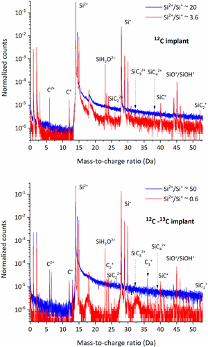
Atom probe tomography was employed to observe and derive the composition of carbon clusters in implanted silicon. This value, which is of interest to the microelectronic industry when considering ion implantation defects, was estimated not to exceed 2 at%. This measurement has been done by fitting the distribution of first nearest neighbor distances between monoatomic carbon ions (C+ and C2+). Carbon quantification has been considerably improved through the detection of molecular ions, using lower electric field conditions as well as equal proportions of 12C and 13C. In these conditions and using another quantification method, we have shown that the carbon content in clusters approaches 50 at%. This result very likely indicates that clusters are nuclei of the SiC phase.

Type
Development and Computation
Copyright
Copyright © The Author(s), 2021. Published by Cambridge University Press on behalf of the Microscopy Society of America

Access options

Get access to the full version of this content by using one of the access options below. (Log in options will check for institutional or personal access. Content may require purchase if you do not have access.)

References

Bazizi, EM, Fazzini, PF, Zechner, C, Tsibizov, A, Kheyrandish, H, Pakfar, A & Cristiano, F (2008). Modelling of boron trapping at end-of-range defects in pre-amorphized ultra-shallow junctions. Mater Sci Eng: B 154, 275278.CrossRefGoogle Scholar
Blavette, D, Vurpillot, F, Pareige, P & Menand, A (2001). A model accounting for spatial overlaps in 3D atom-probe microscopy. Ultramicroscopy 89(1–3), 145153.CrossRefGoogle Scholar
Cacciato, A, Klappe, JGE, Cowern, NEB, Vandervost, W, Biro, LP, Custer, JS & Saris, FW (1996). Dislocation formation and B transient diffusion in C coimplanted Si. J Appl Phys 79(5), 23142325.CrossRefGoogle Scholar
Claverie, A, Colombeau, B, De Mauduit, B, Bonafos, C, Hebras, X, Assayag, GB & Cristiano, F (2003). Extended defects in shallow implants. Appl Phys A 76(7), 10251033.CrossRefGoogle Scholar
Cristiano, F, Bonafos, C, Nejim, A, Lombardo, S, Omri, M, Alquier, D & Claverie, A (1997). Interstitial trapping efficiency of C+ implanted into preamorphised silicon—Control of EOR defects. Nucl Instrum Methods Phys Res B 127, 2226.CrossRefGoogle Scholar
Deconihout, B, Vurpillot, F, Gault, B, Da Costa, G, Bouet, M, Bostel, A & Brunel, M (2007). Toward a laser assisted wide-angle tomographic atom-probe. Surf Interface Anal 39(2–3), 278282.CrossRefGoogle Scholar
Duguay, S, Philippe, T, Cristiano, F & Blavette, D (2010). Direct imaging of boron segregation to extended defects in silicon. Appl Phys Lett 97(24), 242104.CrossRefGoogle Scholar
Dumas, P, Duguay, S, Borrel, J, Gauthier, A, Ghegin, E & Blavette, D (2019). 3D atomic-scale investigation of carbon segregation in phosphorus co-implanted silicon. Appl Phys Lett 115(13), 132103.CrossRefGoogle Scholar
Dumas, P, Duguay, S, Borrel, J, Hilario, F & Blavette, D (2021). Atom probe tomography quantification of carbon in silicon. Ultramicroscopy 220, 113153.CrossRefGoogle ScholarPubMed
Goesele, U, Laveant, P, Scholz, R, Engler, N & Werner, P (1999). Diffusion engineering by carbon in silicon. MRS Online Proc Libr 610(1), 7117112.CrossRefGoogle Scholar
Meisenkothen, F, Steel, EB, Prosa, TJ, Henry, KT & Kolli, RP (2015). Effects of detector dead-time on quantitative analyses involving boron and multi-hit detection events in atom probe tomography. Ultramicroscopy 159, 101111.CrossRefGoogle ScholarPubMed
Peng, Z, Vurpillot, F, Choi, PP, Li, Y, Raabe, D & Gault, B (2018). On the detection of multiple events in atom probe tomography. Ultramicroscopy 189, 5460.CrossRefGoogle ScholarPubMed
Peng, Z, Zanuttini, D, Gervais, B, Jacquet, E, Blum, I, Choi, PP & Gault, B (2019). Unraveling the metastability of Cn2+(n=2–4) clusters. J Phys Chem Lett 10(3), 581588.CrossRefGoogle Scholar
Philippe, T, De Geuser, F, Duguay, S, Lefebvre, W, Cojocaru-Mirédin, O, Da Costa, G & Blavette, D (2009). Clustering and nearest neighbour distances in atom-probe tomography. Ultramicroscopy 109(10), 13041309.CrossRefGoogle ScholarPubMed
Shimizu, Y, Takamizawa, H, Inoue, K, Yano, F, Kudo, S, Nishida, A & Nagai, Y (2016). Impact of carbon co-implantation on boron distribution and activation in silicon studied by atom probe tomography and spreading resistance measurements. Jpn J Appl Phys 55(2), 026501.CrossRefGoogle Scholar
Timans, P, Gelpey, J, McCoy, S, Lerch, W & Paul, S (2006). Millisecond annealing: Past, present and future. MRS Online Proc Libr (OPL) 912, 3.Google Scholar
Vaumousse, D, Cerezo, A & Warren, PJ (2003). A procedure for quantification of precipitate microstructures from three-dimensional atom probe data. Ultramicroscopy 95, 215221.CrossRefGoogle ScholarPubMed