Hostname: page-component-7bb8b95d7b-5mhkq Total loading time: 0 Render date: 2024-10-01T16:14:04.632Z Has data issue: false hasContentIssue false

3D Visualization of Structural and Protein Compositional Changes in Plakophilin-2 Deficient Hearts

Published online by Cambridge University Press:  22 July 2022

Feng-Xia Liang*
Affiliation:
Microscopy Core Laboratory, DART, New York University Grossman School of Medicine, New York, USA
Chantal J.M. van Opbergen
Affiliation:
The Leon H. Charney Division of Cardiology, New York University Grossman School of Medicine, New York, USA
Chris Petzold
Affiliation:
Microscopy Core Laboratory, DART, New York University Grossman School of Medicine, New York, USA
Joseph Sall
Affiliation:
Microscopy Core Laboratory, DART, New York University Grossman School of Medicine, New York, USA
Mario Delmar
Affiliation:
The Leon H. Charney Division of Cardiology, New York University Grossman School of Medicine, New York, USA
*
*Corresponding author: [email protected]

Abstract

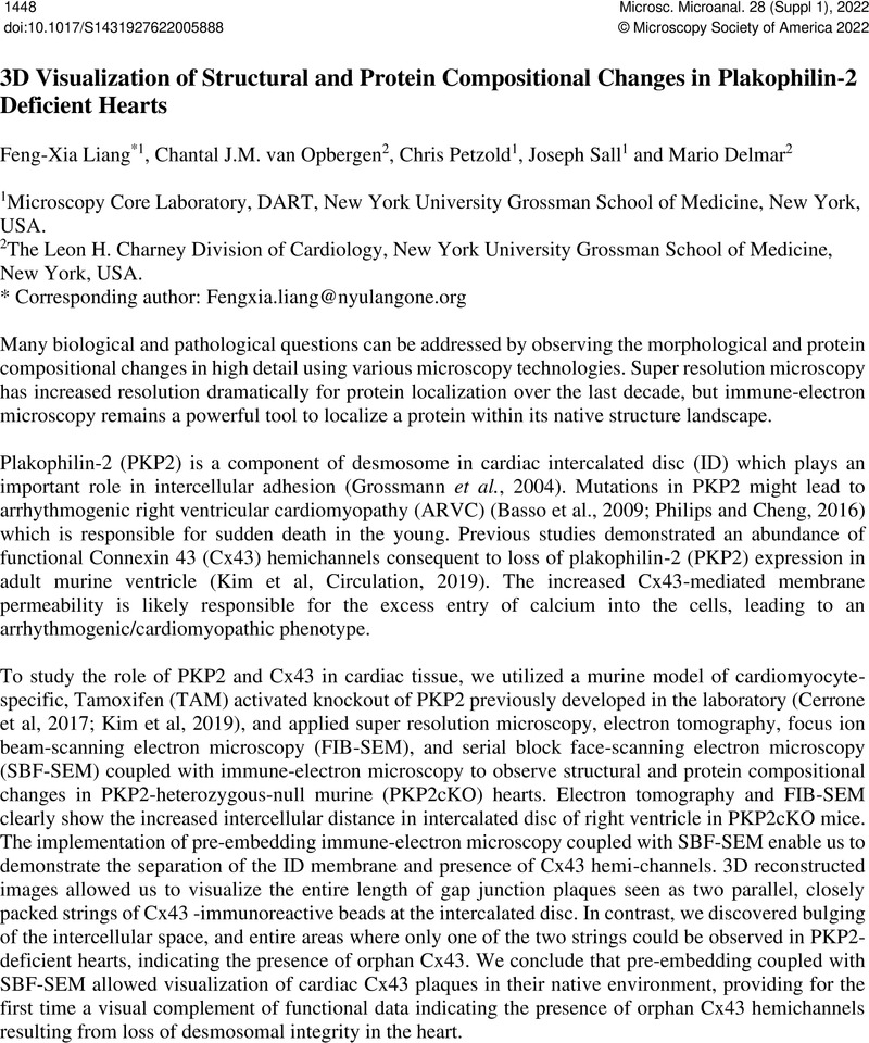
Image of the first page of this content. For PDF version, please use the ‘Save PDF’ preceeding this image.'
Type
3D Volume Electron Microscopy in Biology Research
Copyright
Copyright © Microscopy Society of America 2022

References

Grossman, K.S., et al. , J. Cell Biol., (2004), 167, p.149.CrossRefGoogle Scholar
Basso, C. et al. , Euro. Heart J., (2006), 27 p.1847.CrossRefGoogle Scholar
Philips, B., et al. , Curr. Opin. Cardiol., (2016), 31, p.46.CrossRefGoogle Scholar
Cerrone, M., et al. , Nat. Commun., (2017), 8, p.106.CrossRefGoogle Scholar
Kim, J.C. et al. , Circulation, (2019), 140, p. 1015.Google Scholar