Hostname: page-component-586b7cd67f-2plfb Total loading time: 0 Render date: 2024-11-23T18:48:20.505Z Has data issue: false hasContentIssue false

3D Structure of the Tail Complex of E. coli Bacteriophage DT57C

Published online by Cambridge University Press:  22 July 2022

Rafael Ayala
Affiliation:
Molecular Cryo-Electron Microscopy Unit, Okinawa Institute of Science and Technology Graduate University, Onna-son, Okinawa, Japan
Evgeny Kulikov
Affiliation:
Winogradsky Institute of Microbiology, Research Center of Biotechnology of the Russian Academy of Sciences, Moscow, Russia
Alla Golomidova
Affiliation:
Winogradsky Institute of Microbiology, Research Center of Biotechnology of the Russian Academy of Sciences, Moscow, Russia
Andrey Moiseenko
Affiliation:
Department of Biology, Moscow Lomonosov University, Moscow, Russia N.N.Semenov Federal Research Center for Chemical Physics, Russian Academy of Sciences, Moscow, Russia
Matthias Wolf
Affiliation:
Molecular Cryo-Electron Microscopy Unit, Okinawa Institute of Science and Technology Graduate University, Onna-son, Okinawa, Japan
Andrey V. Letarov
Affiliation:
Winogradsky Institute of Microbiology, Research Center of Biotechnology of the Russian Academy of Sciences, Moscow, Russia Department of Biology, Moscow Lomonosov University, Moscow, Russia
Olga S. Sokolova*
Affiliation:
Department of Biology, Moscow Lomonosov University, Moscow, Russia
*
*Corresponding author: [email protected]

Abstract

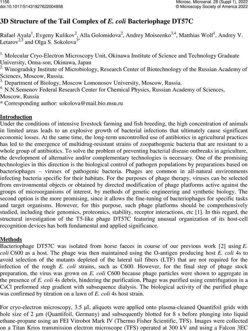
Image of the first page of this content. For PDF version, please use the ‘Save PDF’ preceeding this image.'
Type
On Demand - 3D Structures: From Macromolecular Assemblies to Whole Cells (3DEM FIG)
Copyright
Copyright © Microscopy Society of America 2022

References

Luong, T., Salabarria, A.-C., Roach, D.R. Phage Therapy in the Resistance Era: Where Do We Stand and Where Are We Going? Clin Ther 42(9) (2020), p.1659-1680. doi: 10.1016/j.clinthera.2020.07.014.CrossRefGoogle ScholarPubMed
Golomidova, A., Kulikov, E., Isaeva, A., Manykin, A., Letarov, A. The diversity of coliphages and coliforms in horse feces reveals a complex pattern of ecological interactions. Appl Environ Microbiol, 73 (2007), p.59755981. doi: 10.1128/AEM.01145-07.Google ScholarPubMed
Punjani, A., Rubinstein, J.L., Fleet, D.J., Brubaker, M.A. cryoSPARC: algorithms for rapid unsupervised cryo-EM structure determination. Nat Methods, 14(3) (2017), p. 290-296, doi: 10.1038/nmeth.4169.CrossRefGoogle ScholarPubMed
Scheres, S.H.W. RELION: Implementation of a Bayesian approach to cryo-EM structure determination. J Struct Biol,180(3) (2012), p. 519-530, doi: 10.1016/j.jsb.2012.09.006.CrossRefGoogle ScholarPubMed
Rohou, A., Grigorieff, N. CTFFIND4: Fast and accurate defocus estimation from electron micrographs. J Struct Biol, 192(2) (2015), p. 216-221, doi: 10.1016/j.jsb.2015.08.008.CrossRefGoogle ScholarPubMed
Huet, A, Duda, RL, Boulanger, P, Conway, JF. Capsid expansion of bacteriophage T5 revealed by high resolution cryoelectron microscopy. Proc Natl Acad Sci U S A. 116(42) (2019), p. 21037-21046. doi: 10.1073/pnas.1909645116.CrossRefGoogle ScholarPubMed
Golomidova, AK, Kulikov, EE, Prokhorov, NS, Guerrero-Ferreira, , Knirel, YA, Kostryukova, ES, Tarasyan, KK, Letarov, AV. Branched Lateral Tail Fiber Organization in T5-Like Bacteriophages DT57C and DT571/2 is Revealed by Genetic and Functional Analysis. Viruses, 8(1) (2016), p.26. doi: 10.3390/v8010026.CrossRefGoogle ScholarPubMed
The authors acknowledge funding from the Strategic International Collaborative Research project promoted by Russian Science Foundation (21-44-07002 to OSS) and the Ministry of Agriculture, Forestry and Fisheries, Tokyo, Japan (JP008837 to MW and RA). We acknowledge the OIST imaging section (IMG) for use of the electron microscopy facilityGoogle Scholar