


The "situs" is defined by the location of the heart’s atrial and other viscera. Situs solitus is the position that normally exists, and situs inversus is the opposite of situs solitus, with the morphologically right atrium on the left and the morphologically left atrium on the right. Situs inversus totalis is the medical word for situs inversus when it coexists with dextrocardia. With an incidence of 1:10,000 in some populations, Reference Rashkind and Miller1 it is a rare condition. Major visceral organs are also mirror images of normal in this condition. Five to ten percent of these patients also have CHD, of which transposition of great arteries is the most common.
Case report
A term newborn girl who is 3300 gr and 21 days old was referred to our hospital from an external hospital. She was intubated because the patient was desaturated and a prostaglandin infusion was initiated.
Echocardiography showed situs inversus with dextrocardia, concordant atrio-ventricular connections, and discordant ventriculo-arterial connections. Aorta was anterior to the left of pulmonary artery with usual coronary pattern for dextrocardia (Fig. 1), a non-restrictive atrial septal defect, thin patent ductus arteriosus, and left ventriküler outflow obstruction without ventricular septal defect (Fig. 2). Left ventricle was very hypertrophied. Also abdominal ultrasound demonstrated the stomach and spleen on the right side and the liver on the left.
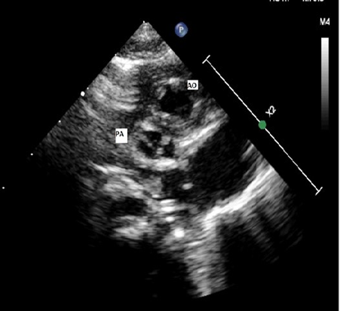
Figure 1. The relationship between aorta and pulmonary artery.

Figure 2. The measurment of LVOTO.
On physical examination, child was cyanosed with baseline oxygen saturation of 58% with respiratory distress with respiratory rates of 70 per minute and subcostal retractions. She had normal precordium on palpation with normal first heart sound and a single loud second heart sound. Despite medical treatment, her general condition worsened. In her arterial blood gas test, her oxygen saturation was low and she had got lactate elevation. We decided to perform PDA stent implantation. Patient underwent PDA stent implantation under emergency conditions. After successful catheterisation procedure, the patient’s arterial blood gas results improved to 85%. She was extubated within 10 days later and weaned from haemodynamic support. After post-procedure echocardiography, we noticed that the patient’s pulmonary stenosis gradient decreased. Her left ventricular outflow tract stenosis is dynamic stenosis due to left ventricular hypertrophy and also due to aneurysm tissues closing VSD. It was decided that left ventricle outflow tract obstruction could be removed surgically, probably due to the aneurysm tissues closing the VSD and we decided to perform arteriel switch operation.
On her 46th day, the patient underwent arterial switch operation and resection of left ventricular outflow tract obstruction. Under full-flow cardiopulmonary bypass, the aorta and pulmonary artery were transected, the stent placed in the PDA was removed, LeCompte manoeuvre was performed, and the coronary arteries were re-anastomosed to the aorta in the standard fashion. During the examination of perimembranous region and left ventricular outflow tract that subpulmonic VSD was closed by tricuspid leaflets and it causes left ventricle outflow tract obstruction. Left ventricle outflow tract obstruction forming septal structures was cleaned. Post-operative transesophageal echocardiography demonstrated no new obstruction in the neo-aorta or neo-pulmonary artery, and no residual shunting at the atrial or ventricular level. The patient returned from the operating room on milrinone (0.5 mcg/kg/min) and ventilation settings. After uneventful follow-up, she was extubated on post-operative second day. On post-operative day 10, she was discharged on a good condition by recommendation for control in the paediatric cardiology clinic.
Discussion
Situs inversus totalis is a rare condition with a prevalence of 1:10,000 in some populations. Reference Rashkind and Miller1 About 5–10 % of patients with this defect also have CHD. D-transposition of the great arteries is the most common congenital heart lesion which affects about 2.6–7.8% of these patients. Reference Castañeda, Norwood, Jonas, Colon, Sanders and Lang2,Reference Neches, Park, Ettedgui, Garson, Bricker, Fisher and Neish3 Surgical management of D-transposition of the great arteries has evolved over several decades under the influence of a few key surgical pioneers. Reference Blalock and Hanlon4,Reference Senning5 The arterial switch operation has become the standard surgical repair for D-transposition of the great arteries in the absence of left ventricular outflow tract obstruction. The current mortality risk for neonatal arterial switch operation is less than 5%, even in the presence of a complex coronary arrangement. Reference Planche, Lacour-Gayet and Serraf6
In addition to the situs inversus, dextrocardia, transposition of the great arteries, there was left ventricle outflow tract obstruction with intact ventricular septum in echocardiographic examination of our patient. The position and size of VSD and the morphology of left ventricle outflow tract obstruction determine surgical treatment in these patients. If left ventricle outflow tract obstruction is not resectable, total repair is possible only with committed and non-restrictive VSD. While left ventricle outflow tract obstruction is seen 30–35% in D-transposition of the great arteries, its presence with intact ventricular septum is very rare as 0.7%. Reference Wernovsky, Allen, Driscoll, Shaddy and Feltes7 The obstruction may be dynamic or an atomic which can be from the subvalvar fibrous tissue tags, hypertrophied band or valvar stenosis. In our patient, the aneurysm tissues that closed the VSD caused left ventricle outflow tract obstruction. It is known that these patients are very cyanotic early in life and these patients need a non-restrictive interatrial communication and sometimes systemic-pulmonary artery shunt. Our patient’s atrial septal defect was non-restrictive and we preferred to perform an emergency patent ductus arteriosus stent because of thin patent ductus arteriosus. Her general condition improved after emergency patent ductus arteriosus stent implantation but after this procedure her left ventricle outflow tract obstruction gradient decreased. We decided to perform arteriel switch because we thought that left ventricle outflow tract obstruction tissue was resectable. Her aortic annulus 12 mm (z skor: 5,42) and pulmonary annulus 7.6 mm (z skor: −0.29) so we have to do left ventricle outflow tract obstruction resection and ASO with VSD closure. Reference Hazekamp, Portela and Bartelings8,Reference Honjo, Kotani and Bharucha9 We have reported of successful arterial switch, left ventricle outflow tract obstruction resection in a patient with atrial and visceral situs in-versus totalis, dextrocardia, atrioventricular concordance, and ventriculo-arterial discordance (I,L,L).
To our knowledge, this is a very rare cardiac anatomy in the literature. However, transposition of the great arteries is more frequent in situs inversus than in situs solitus. Reference Unolt, Putotto and Silvestri10 When we look at the previous another reports, we have observed a case about and a situs inversus with dextrocardia and D-transposition of the great arteries with intact ventricular septum. Reference Awasthy, Kapoor, Upreti and Dagar11
In this case report, we discussed an extremely rare patient with dextrocardic transposition of the great arteries and intact ventricular septum. We performed successful PDA stent implantation followed by neonatal arterial switch and left ventricle outflow tract obstruction resection in a patient with atrioventricular concordance and ventriculo-arterial discordance.
Acknowledgements
None.
Competing interests
None.


