Skip to main content Accessibility help
×
Hostname: page-component-586b7cd67f-2brh9 Total loading time: 0 Render date: 2024-11-29T01:32:10.442Z Has data issue: false hasContentIssue false

Chapter 36 - Forensic Pathology Aspects of Sudden Unexpected Death in Infancy and Childhood

from Section 8 - SUDI/SUID Which is Not SIDS

Published online by Cambridge University Press:  04 June 2019

Marta C. Cohen
Affiliation:
Sheffield Children’s Hospital
Irene B. Scheimberg
Affiliation:
Royal London Hospital
J. Bruce Beckwith
Affiliation:
Loma Linda University School of Medicine
Fern R. Hauck
Affiliation:
University of Virginia
Get access

Summary

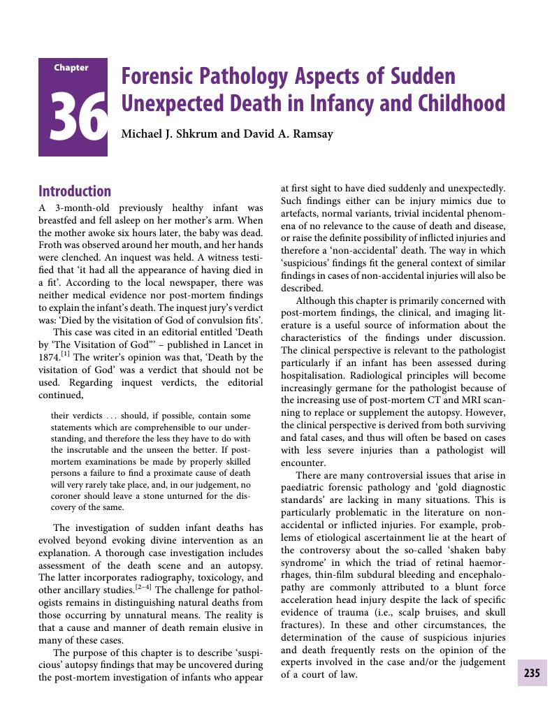
Image of the first page of this content. For PDF version, please use the ‘Save PDF’ preceeding this image.'
Type
Chapter
Information
Publisher: Cambridge University Press
Print publication year: 2019

Access options

Get access to the full version of this content by using one of the access options below. (Log in options will check for institutional or personal access. Content may require purchase if you do not have access.)

References

Editorial. Death by ‘The Visitation of God’. Lancet, 1874; 1:452.Google Scholar
Corey, TS, Hanzlick, R, Howard, J, et al. NAME Ad Hoc Committee on Sudden Unexplained Infant Death. A functional approach to sudden unexplained infant deaths. Am J Forensic Med Pathol, 2007; 28:271–7.Google Scholar
Krous, HF, Beckwith, JB, Byard, RW, et al. Sudden Infant Death Syndrome and unclassified sudden infant deaths: a definitional and diagnostic approach. Pediatrics, 2004; 114:234–8.CrossRefGoogle ScholarPubMed
Shapiro-Mendoza, CK, Camperlengo, L, Ludvigsen, R, et al. Classification system for the Sudden Unexpected Infant Death Case Registry and its application. Pediatrics, 2014; 134:e21019.CrossRefGoogle ScholarPubMed
Matshes, EW, Lew, EO. An approach to the classification of apparent asphyxial infant deaths. Acad Forensic Pathol, 2017; 7:200–11.CrossRefGoogle Scholar
Hanzlick, R. Pulmonary hemorrhage in deceased infants: baseline data for further study of infant mortality. Am J Forensic Med Pathol, 2001; 22:188–92.CrossRefGoogle ScholarPubMed
Willinger, M, James, LS, Catz, C. Defining the Sudden Infant Death Syndrome (SIDS): deliberations of an expert panel convened by the National Institute of Child Health and Human Development. Pediatr Pathol, 1991; 11:677–84.CrossRefGoogle Scholar
Goudge, ST. Inquiry into Pediatric Forensic Pathology in Ontario Report. Toronto: Queen’s Printer for Ontario, Ontario Ministry of the Attorney General, 2008.Google Scholar
Collins, KA. Death by overlaying and wedging: a 15-year retrospective study. Am J Forensic Med Pathol, 2001; 22:155–9.Google Scholar
Hicks, LJ, Scanlon, MJ, Bostwick, TC, et al. Death by smothering and its investigation. Am J Forensic Med Pathol, 1990; 11:291–3.CrossRefGoogle ScholarPubMed
Leduc, D. Well Beings: A Guide to Health in Child Care, 3rd edn (revised). Ottawa: Canadian Paediatric Society, 2015.Google Scholar
Tieder, JS, Bonkowsky, JL, Etzel, RA, et al. Clinical practice guideline: brief resolved unexplained events (formerly apparent life-threatening events) and evaluation of lower-risk infants: executive summary. Pediatrics, 2016; 137(5):e20160591; http://pediatrics.aappublications.org/content/pediatrics/137/5/e20160590.full.pdf (accessed 31 October 2018).CrossRefGoogle ScholarPubMed
Byard, RW, Stewart, WA, Telfer, S, et al. Assessment of pulmonary and intrathymic hemosiderin deposition in Sudden Infant Death Syndrome. Pediatr Pathol Lab Med, 1997; 17:275–82.CrossRefGoogle ScholarPubMed
Jackson, CM, Gilliland, MG. Frequency of pulmonary hemosiderosis in Eastern North Carolina. Am J Forensic Med Pathol, 2000; 21:36–8.Google Scholar
Kempe, CH, Silverman, FN, Steele, BF, et al. The battered-child syndrome. JAMA, 1962; 181:1724.CrossRefGoogle ScholarPubMed
Memorandum #63. New Protocol to be Used in the Investigation of the Sudden and Unexpected Death of Any Child Under 2 Years of Age. Toronto: Office of the Chief Coroner of Ontario, 1995.Google Scholar
DiMaio, VJM, DiMaio, DJ. Forensic Pathology. New York: Elsevier, 1989.Google Scholar
Pinneri, K, Matshes, E. Recommendations for the autopsy of an infant who has died suddenly and unexpectedly. Acad Forensic Pathol, 2017; 7:171–81.CrossRefGoogle ScholarPubMed
Shkrum, MJ, Ramsay, DA. Forensic Pathology of Trauma: Common Problems for the Pathologist. Totowa, NJ: Humana Press, 2007.CrossRefGoogle Scholar
Byard, RW. Unexpected infant death: lessons from the Sally Clark case. Med J Aust, 2004; 181:52–4.CrossRefGoogle ScholarPubMed
Green, FC. Child abuse and neglect: a priority problem for the private physician. Pediatr Clin North Am, 1975; 22:329–39.CrossRefGoogle ScholarPubMed
Maguire, S, Mann, MK, Sibert, J, et al. Are there patterns of bruising in childhood which are diagnostic or suggestive of abuse? A systematic review. Arch Dis Child, 2005; 90:182–6.Google Scholar
Patel, B, Butterfield, R. Common skin and bleeding disorders that can potentially masquerade as child abuse. Am J Med Genet C Semin Med Genet, 2015; 169:328–36.CrossRefGoogle ScholarPubMed
Schwartz, KA, Metz, J, Feldman, K, et al. Cutaneous findings mistaken for physical abuse: present but not pervasive. Pediatr Dermatol, 2014; 31:146–55.CrossRefGoogle Scholar
Tsokos, M. Diagnostic criteria for cutaneous injuries in child abuse: classification, findings, and interpretation. Forensic Sci Med Pathol, 2015; 11:235–42.CrossRefGoogle ScholarPubMed
Anderst, JD, Carpenter, SL, Abshire, TC. Evaluation for bleeding disorders in suspected child abuse. Pediatrics, 2013; 131:e131422.Google Scholar
Pierce, MC, Kaczor, K, Aldridge, S, et al. Bruising characteristics discriminating physical child abuse from accidental trauma. Pediatrics, 2010; 125:6774.Google Scholar
Maguire, S, Mann, MK, Sibert, J, et al. Can you age bruises accurately in children? A systematic review. Arch Dis Child, 2005; 90:187–9.Google Scholar
Bariciak, ED, Plint, AC, Gaboury, I, et al. Dating of bruises in children: an assessment of physician accuracy. Pediatrics, 2003; 112:804–7.CrossRefGoogle ScholarPubMed
Vanezis, P. Interpreting bruises at necropsy. J Clin Pathol, 2001; 54:348–55.CrossRefGoogle ScholarPubMed
Nichols, GR, Corey, TS, Davis, GJ. Nonfracture-associated fatal fat embolism in a case of child abuse. J Forensic Sci, 1990; 35:493–9.Google Scholar
Metz, JB, Schwartz, KA, Feldman, KW, et al. Non-cutaneous conditions clinicians might mistake for abuse. Arch Dis Child, 2014; 99:817–23.CrossRefGoogle ScholarPubMed
Asati, DP, Singh, S, Sharma, VK, et al. Dermatoses misdiagnosed as deliberate injuries. Med Sci Law, 2012; 52:198204.CrossRefGoogle ScholarPubMed
Carpenter, SL, Abshire, TC, Anderst, JD. Evaluating for suspected child abuse: conditions that predispose to bleeding. Pediatrics, 2013; 131:e135773.Google Scholar
Chester, DL, Jose, RM, Aldlyami, E, et al. Non-accidental burns in children – are we neglecting neglect? Burns J Int Soc Burn Inj, 2006; 32:222–8.Google Scholar
Lilly, E, Kundu, RV. Dermatoses secondary to Asian cultural practices. Int J Dermatol, 2012; 51:372–9; quiz 379–82.Google Scholar
Pollanen, MS, Smith, CR, Chiasson, DA, et al. Fatal child abuse-maltreatment syndrome. A retrospective study in Ontario, Canada, 1990–1995. Forensic Sci Int, 2002; 126:101–4.Google ScholarPubMed
Pelletti, G, Tambuscio, S, Montisci, M, et al. Misinterpretation of anogenital findings and misdiagnosis of child sexual abuse: The role of the forensic pathologist. J Pediatr Adolesc Gynecol, 2016; 29:e2931.Google Scholar
McCann, J, Reay, D, Siebert, J, et al. Postmortem perianal findings in children. Am J Forensic Med Pathol, 1996; 17:289–98.CrossRefGoogle ScholarPubMed
Hobbs, CJ, Wright, CM. Anal signs of child sexual abuse: a case-control study. BMC Pediatr, 2014; 14:128.CrossRefGoogle ScholarPubMed
Ross, AH, Juarez, CA. Skeletal and radiological manifestations of child abuse: Implications for study in past populations. Clin Anat NYN, 2016; 29:844–53.Google Scholar
Akbarnia, B, Torg, JS, Kirkpatrick, J, et al. Manifestations of the battered-child syndrome. J Bone Joint Surg Am, 1974; 56:1159–66.Google Scholar
Merten, DF, Radkowski, MA, Leonidas, JC. The abused child: a radiological reappraisal. Radiology, 1983; 146:377–81.CrossRefGoogle Scholar
Conway, JJ, Collins, M, Tanz, RR, et al. The role of bone scintigraphy in detecting child abuse. Semin Nucl Med, 1993; 23:321–33.CrossRefGoogle ScholarPubMed
Brogdon, BG (Byron, G, Shwayder, T, Elifritz, J. Child Abuse and its Mimics in Skin and Bone. Boca Raton, FL: CRC Press, 2012.CrossRefGoogle Scholar
Klotzbach, H, Delling, G, Richter, E, et al. Post-mortem diagnosis and age estimation of infants’ fractures. Int J Legal Med, 2003; 117:82–9.CrossRefGoogle ScholarPubMed
Sanchez, TR, Grasparil, AD, Chaudhari, R, et al. Characteristics of rib fractures in child abuse – The role of low-dose chest computed tomography. Pediatr Emerg Care, 2018; 34(2):81–3.Google Scholar
Maguire, S, Mann, M, John, N, et al. Does cardiopulmonary resuscitation cause rib fractures in children? A systematic review. Child Abuse Negl, 2006; 30:739–51.CrossRefGoogle ScholarPubMed
Kleinman, PK, Marks, SC, Nimkin, K, et al. Rib fractures in 31 abused infants: postmortem radiologic-histopathologic study. Radiology, 1996; 200:807–10.Google Scholar
Lonergan, GJ, Baker, AM, Morey, MK, et al. From the archives of the AFIP. Child abuse: radiologic-pathologic correlation. Radiogr Rev Publ Radiol Soc N Am Inc, 2003; 23:811–45.Google ScholarPubMed
Carty, HM. Fractures caused by child abuse. J Bone Joint Surg Br, 1993; 75:849–57.Google ScholarPubMed
Sanchez, TR, Nguyen, H, Palacios, W, et al. Retrospective evaluation and dating of non-accidental rib fractures in infants. Clin Radiol, 2013; 68:e46771.CrossRefGoogle ScholarPubMed
Chapman, S. The radiological dating of injuries. Arch Dis Child, 1992; 67:1063–5.CrossRefGoogle ScholarPubMed
Prosser, I, Lawson, Z, Evans, A, et al. A timetable for the radiologic features of fracture healing in young children. AJR Am J Roentgenol, 2012; 198:1014–20.CrossRefGoogle ScholarPubMed
Betz, P, Liebhardt, E. Rib fractures in children – resuscitation or child abuse? Int J Legal Med, 1994; 106:215–18.Google Scholar
Weber, MA, Risdon, RA, Offiah, AC, et al. Rib fractures identified at post-mortem examination in sudden unexpected deaths in infancy (SUDI). Forensic Sci Int, 2009; 189:7581.CrossRefGoogle ScholarPubMed
Kogutt, MS, Swischuk, LE, Fagan, CJ. Patterns of injury and significance of uncommon fractures in the battered child syndrome. Am J Roentgenol Radium Ther Nucl Med, 1974; 121:143–9.CrossRefGoogle ScholarPubMed
Duval, JV, Andrew, TA. Two thumb method of infant CPR: Is there an increased risk for posterior rib fractures? Paper presented at National Association of Medical Examiners 41st Annual Meeting, 2007, Savannah, Georgia.Google Scholar
Clouse, JR, Lantz, PE. Posterior rib fractures in infants associated with cardiopulmonary resuscitation. Paper presented at American Academy of Forensic Sciences Annual Meeting, 2008, Washington, DC.Google Scholar
Matshes, EW, Lew, EO. Two-handed cardiopulmonary resuscitation can cause rib fractures in infants. Am J Forensic Med Pathol, 2010; 31:303–7.CrossRefGoogle ScholarPubMed
van Rijn, RR, Bilo, RAC, Robben, SGF. Birth-related mid-posterior rib fractures in neonates: a report of three cases (and a possible fourth case) and a review of the literature. Pediatr Radiol, 2009; 39:30–4.CrossRefGoogle Scholar
Pandya, NK, Baldwin, K, Kamath, AF, et al. Unexplained fractures: child abuse or bone disease? A systematic review. Clin Orthop, 2011; 469:805–12.CrossRefGoogle ScholarPubMed
Cannell, JJ, Holick, MF. Multiple unexplained fractures in infants and child physical abuse. J Steroid Biochem Mol Biol, 2018; 175:1822.Google Scholar
Miller, M, Mirkin, LD. Classical metaphyseal lesions thought to be pathognomonic of child abuse are often artifacts or indicative of metabolic bone disease. Med Hypotheses, 2018; 115:6571.CrossRefGoogle ScholarPubMed
Leavitt, EB, Pincus, RL, Bukachevsky, R. Otolaryngologic manifestations of child abuse. Arch Otolaryngol Head Neck Surg, 1992; 118:629–31.CrossRefGoogle ScholarPubMed
Phillips, VM, Van Der Heyde Y. Oro-facial trauma in child abuse fatalities. South Afr Med J Suid-Afr Tydskr Vir Geneeskd, 2006; 96:213–15.Google ScholarPubMed
Maguire, SA, Upadhyaya, M, Evans, A, et al. A systematic review of abusive visceral injuries in childhood – their range and recognition. Child Abuse Negl, 2013; 37:430–45.Google Scholar
Plunkett, J. Resuscitation injuries complicating the interpretation of premortem trauma and natural disease in children. J Forensic Sci, 2006; 51:127–30.Google Scholar
Kleinman, PK, Raptopoulos, VD, Brill, PW. Occult nonskeletal trauma in the battered-child syndrome. Radiology, 1981; 141:393–6.Google Scholar
Huntimer, CM, Muret-Wagstaff, S, Leland, NL. Can falls on stairs result in small intestine perforations? Pediatrics, 2000; 106:301–5.Google Scholar
Barnes, PM, Norton, CM, Dunstan, FD, et al. Abdominal injury due to child abuse. Lancet, 2005; 366:234–5.CrossRefGoogle ScholarPubMed
Davison, AM, Lazda, EJ. Small bowel perforation and fatal peritonitis following a fall in a 21-month-old child. Forensic Sci Med Pathol, 2008; 4:250–4.CrossRefGoogle Scholar
Gaines, BA, Shultz, BS, Morrison, K, et al. Duodenal injuries in children: beware of child abuse. J Pediatr Surg, 2004; 39:600–2.Google Scholar
Nimkin, K, Teeger, S, Wallach, MT, et al. Adrenal hemorrhage in abused children: imaging and postmortem findings. AJR Am J Roentgenol, 1994; 162:661–3.Google Scholar
Matshes, EW, Lew, EO. Do resuscitation-related injuries kill infants and children? Am J Forensic Med Pathol, 2010; 31:178–85.CrossRefGoogle ScholarPubMed
Madea, B, Ortmann, J, Doberentz, E. Forensic aspects of starvation. Forensic Sci Med Pathol, 2016; 12:276–98.Google Scholar
Folkerth, RD Nunez, J, Zhanna, G, McGuone, D. Neuropathologic examination in sudden unexpected deaths in infancy and childhood: recommendations for highest diagnostic yield and cost-effectiveness in forensic settings. Acad Forensic Pathol, 2017; 7:182–99.CrossRefGoogle ScholarPubMed
Mortazavi, MM, Shane Tubbs, R, Riech, S, et al. Anatomy and pathology of the cranial emissary veins: A review with surgical implications. Neurosurgery, 2012; 70:1312–18.Google Scholar
Aliabadi, H, Miller, J, Radnakrishnan, S, et al. Spontaneous intrauterine ‘ping-pong’ fracture: review and case illustration. Neuropediatrics, 2009; 40:73–5.CrossRefGoogle ScholarPubMed
Heise, RH, Srivatsa, PJ, Karsell, PR. Spontaneous intrauterine linear skull fracture: a rare complication of spontaneous vaginal delivery. Obstet Gynecol, 1996; 87:851–4.Google Scholar
Loire, M, Barat, M, Mangyanda Kinkembo, L, et al. Spontaneous ping-pong parietal fracture in a newborn. Arch Dis Child Fetal Neonatal Ed, 2017; 102:F160F161.Google Scholar
Idriz, S, Patel, JH, Ameli Renani, S, et al. CT of normal developmental and variant anatomy of the pediatric skull: Distinguishing trauma from normality. RadioGraphics, 2015; 35:1585–601.Google Scholar
Sanchez, T, Stewart, D, Walvick, M, et al. Skull fracture vs. accessory sutures: How can we tell the difference? Emerg Radiol, 2010; 17:413–18.CrossRefGoogle ScholarPubMed
Marti, B, Sirinelli, D, Maurin, L, et al. Wormian bones in a general paediatric population. Diagn Interv Imaging, 2013; 94:428–32.Google Scholar
Bellary, SS, Steinberg, A, Mirzayan, N, et al. Wormian bones: A review. Clin Anat, 2013; 26:922–7.CrossRefGoogle ScholarPubMed
Droms, RJ, Rork, JF, McLean, R, et al. Menkes disease mimicking child abuse. Pediatr Dermatol, 2017; 34: e132e134.Google Scholar
Wiedijk, JEF, Soerdjbalie-Maikoe, V, Maat, GJR, et al. An accessory skull suture mimicking a skull fracture. Forensic Sci Int, 2016; 260:e1113.CrossRefGoogle ScholarPubMed
Plunkett, J. Fatal pediatric head injuries caused by short-distance falls. Am J Forensic Med Pathol, 2001; 22:112.Google Scholar
Reiber, GD. Fatal falls in childhood. How far must children fall to sustain fatal head injury? Report of cases and review of the literature. Am J Forensic Med Pathol, 1993; 14:201–7.CrossRefGoogle ScholarPubMed
Van Ee, C, Moroski-Browne, B, Raymond, D, et al. Evaluation and refinement of the CRABI-6 anthropomorphic test device injury criteria for skull fracture. In: ASME 2009 International Mechanical Engineering Congress and Exposition: vol. 2,Biomedical and Biotechnology Engineering. Lake Buena Vista, FL: ASME Digital Library: 2009:387–93.Google Scholar
Reece, RM, Sege, R. Childhood head injuries: accidental or inflicted? Arch Pediatr Adolesc Med, 2000; 154:1115.Google Scholar
Harwood-Nash, DC, Hendrick, EB, Hudson, AR. The significance of skull fractures in children. A study of 1,187 patients. Radiology, 1971; 101:151–6.Google Scholar
Chadwick, DL, Bertocci, G, Castillo, E, et al. Annual risk of death resulting from short falls among young children: less than 1 in 1 million. Pediatrics, 2008; 121:1213–24.CrossRefGoogle Scholar
Helfer, RE, Slovis, TL, Black, M. Injuries resulting when small children fall out of bed. Pediatrics, 1977; 60:533–5.Google Scholar
Kravitz, H, Driessen, G, Gomberg, R, et al. Accidental falls from elevated surfaces in infants from birth to one year of age. Pediatrics, 1969; 44:869–76.Google Scholar
Squier, W. Shaken baby syndrome: the quest for evidence. Dev Med Child Neurol, 2008; 50:1014.Google Scholar
Huang, MHS, Mouradian, WE, Cohen, SR, et al. The differential diagnosis of abnormal head shapes: Separating craniosynostosis from positional deformities and normal variants. Cleft Palate Craniofac J, 1998; 35:204–11.CrossRefGoogle ScholarPubMed
Rogers, GF. Deformational plagiocephaly, brachycephaly, and scaphocephaly. Part II. J Craniofac Surg, 2011; 22:1723.Google Scholar
Cunningham, ML, Heike, CL. Evaluation of the infant with an abnormal skull shape. Curr Opin Pediatr, 2007; 19:645–51.Google Scholar
Ginelliová, A, Farkaš, D, Iannaccone, SF, et al. Sudden death associated with syndromic craniosynostosis. Forensic Sci Med Pathol, 2016; 12:506–9.CrossRefGoogle ScholarPubMed
Von der Hagen, M, Pivarcsi, M, Liebe, J, et al. Diagnostic approach to microcephaly in childhood: A two-center study and review of the literature. Dev Med Child Neurol, 2014; 56:732–41.Google Scholar
Olney, AH. Macrocephaly Syndromes. Semin Pediatr Neurol, 2007; 14:128–35.CrossRefGoogle ScholarPubMed
Beasley, M. Age of fontanelles/cranial sutures closure. Centre for Academic Research and Training in Anthropogeny: Matrix of Comparitive Anthropogeny https://carta.anthropogeny.org/moca/topics/age-closure-fontanelles-sutures (accessed 31 October 2018).Google Scholar
Kiesler, J, Ricer, R. The abnormal fontanelle. Am Fam Physician, 2003; 67:2547–52.Google Scholar
Pindrik, J, Ye, X, Ji, BG, et al. Anterior fontanelle closure and size in full-term children based on head computed tomography. Clin Pediatr Phil, 2014; 53:1149–57.Google ScholarPubMed
Cohen, MC, Scheimberg, I. Evidence of occurrence of intradural and subdural hemorrhage in the perinatal and neonatal period in the context of hypoxic Ischemic encephalopathy: an observational study from two referral institutions in the United Kingdom. Pediatr Dev Pathol, 2009; 12:169–76.Google Scholar
Scheimberg, I, Cohen, MC, Zapata Vazquez, RE, et al. Nontraumatic intradural and subdural hemorrhage and hypoxic ischemic encephalopathy in fetuses, infants, and children up to three years of age: analysis of two audits of 636 cases from two referral centers in the United Kingdom. Pediatr Dev Pathol, 2013; 16:149–59.Google Scholar
Rutty, GN, Squier, WM. Subdural hematoma in children. In: Essentials of Autopsy Practice: Current Methods and Modern Trends. Berlin: Springer, 2006:131–53.Google Scholar
Pollanen, MS. Subdural hemorrhage in infancy: keep an open mind. Forensic Sci Med Pathol, 2011; 7:298300.Google Scholar
De Leeuw, M, Beuls, E, Jorens, P, et al. Delta-storage pool disease as a mimic of abusive head trauma in a 7-month-old baby: a case report. J Forensic Leg Med, 2013; 20:520–1.CrossRefGoogle Scholar
Ganesh, A, Jenny, C, Geyer, J, et al. Retinal hemorrhages in type I osteogenesis imperfecta after minor trauma. Ophthalmology, 2004; 111:1428–31.Google Scholar
Haddad, MH. Hemorrhages after minor trauma (letter). Ophthalmology, 2005; 112:737–8.Google Scholar
Nassogne, M-C, Sharrard, M, Hertz-Pannier, L, et al. Massive subdural haematomas in Menkes disease mimicking shaken baby syndrome. Childs Nerv Syst ChNS Off J Int Soc Pediatr Neurosurg, 2002; 18:729–31.Google Scholar
Zielonka, M, Braun, K, Bengel, A, et al. Severe acute subdural hemorrhage in a patient with glutaric aciduria type I after minor head trauma: A case report. J Child Neurol, 2015; 30:1065–9.Google Scholar
Cohen, MC, Yap, S, Olpin, E. Inherited metabolic disease and sudden unexpected death. In: Payne-James, J, Byard, RW, eds. Encyclopaedia of Forensic and Legal Medicine, 2nd edn., Oxford: Elsevier, 2016: 8595.CrossRefGoogle Scholar
Christian, CW. The evaluation of suspected child physical abuse. Pediatrics, 2015; 135 e1337e1354.Google Scholar
Lynøe, N, Elinder, G, Hallberg, B, et al. Insufficient evidence for ‘shaken baby syndrome’ – a systematic review. Acta Paediatr, 2017; 2:17.Google Scholar
Rorke-Adams, LB. The triad of retinal haemorrhage, subdural haemorrhage and encephalopathy in an infant unassociated with evidence of physical injury is not the result of shaking, but is most likely to have been caused by a natural disease: No. J Prim Health Care, 2011; 3:161–3.Google Scholar
Saunders, D, Raissaki, M, Servaes, S, et al. Throwing the baby out with the bath water – response to the Swedish Agency for Health Technology Assessment and Assessment of Social Services (SBU) report on traumatic shaking. Pediatr Radiol, 2017; 47(11)1386–9.Google Scholar
Squier, W. The triad of retinal haemorrhage, subdural haemorrhage and encephalopathy in an infant unassociated with evidence of physical injury is not the result of shaking, but is most likely to have been caused by a natural disease: Yes. J Prim Health Care, 2011; 3:159–61.Google Scholar
Squier, W. ‘Shaken baby syndrome’ and forensic pathology. Forensic Sci Med Pathol, 2014; 10:248–50.Google Scholar
Del Bigio, MR, Phillips, SM. Retroocular and subdural hemorrhage or hemosiderin deposits in pediatric autopsies. J Neuropathol Exp Neurol, 2017; 62:513–69.Google Scholar
Rooks, VJ, Eaton, JP, Ruess, L, et al. Prevalence and evolution of intracranial hemorrhage in asymptomatic term infants. Am J Neuroradiol, 2008; 29:1082–9.CrossRefGoogle ScholarPubMed
Kelly, P, Hayman, R, Shekerdemian, LS, et al. Subdural hemorrhage and hypoxia in infants with congenital heart disease. Pediatrics, 2014; 134:e77381.Google Scholar
Watts, P, Maguire, S, Kwok, T, et al. Newborn retinal hemorrhages: A systematic review. J AAPOS, 2013; 17:70–8.CrossRefGoogle ScholarPubMed
Leeuw, MD, Beuls, E, Jorens, PG, et al. The optic nerve sheath hemorrhage is a non-specific finding in cases of suspected child abuse. J Forensic Leg Med, 2015; 36:43–8.Google Scholar
deVeber, G, Andrew, M, Adams, C, et al. Cerebral sinovenous thrombosis in children. N Engl J Med, 2001; 345:417–23.Google Scholar
Hedlund, GL. Cerebral sinovenous thrombosis in pediatric practice. Pediatr Radiol, 2013; 43:173–88.Google Scholar
McLean, LS; Frasier, LD; Hedlund, GL. Does intracranial venous thrombosis cause subdural hemorrhage in the pediatric population? AJNR, 2012; 33:1281–4.Google Scholar
Krasnokutsky, MV. Cerebral venous thrombosis: a potential mimic of primary traumatic brain injury in infants. AJR Am J Roentgenol, 2011; 197:W5037.CrossRefGoogle ScholarPubMed
Squier, W. The ‘shaken baby’ syndrome: pathology and mechanisms. Acta Neuropathol (Berl), 2011; 122:519–42.CrossRefGoogle ScholarPubMed
Sieswerda-Hoogendoorn, T, Soerdjbalie-Maikoe, V, de Bakker, H, et al. Postmortem CT compared to autopsy in children; concordance in a forensic setting. Int J Legal Med, 2014; 128:957–65.Google Scholar
Krous, HF, Haas, EA, Chadwick, AE, et al. Delayed death in Sudden Infant Death Syndrome: a San Diego SIDS/SUDC Research Project 15-year population-based report. Forensic Sci Int, 2008; 176:209–16.Google Scholar
Geddes, JF, Tasker, RC, Hackshaw, AK, et al. Dural haemorrhage in non-traumatic infant deaths: does it explain the bleeding in ‘shaken baby syndrome’? Neuropathol Appl Neurobiol, 2004; 29:1422.Google Scholar
Smith, C, Bell, JE, Keeling, JW, et al. Dural haemorrhage in nontraumatic infant deaths: does it explain the bleeding in ‘shaken baby syndrome’? Geddes JE et al. A response. Neuropathol Appl Neurobiol, 2003; 29:411–12.Google Scholar
Punt, J, Bonshek, RE, Jaspan, T, et al. The ‘unified hypothesis’ of Geddes et al. is not supported by the data. Pediatr Rehabil, 2004; 7:173–84.CrossRefGoogle ScholarPubMed
Byard, RW, Blumbergs, P, Rutty, G, et al. Lack of evidence for a causal relationship between hypoxic-ischemic encephalopathy and subdural hemorrhage in fetal life, infancy, and early childhood. Pediatr Dev Pathol, 2007; 10:348–50.Google Scholar
Hurley, M, Dineen, R, Padfield, CJH, et al. Is there a causal relationship between the hypoxia-ischaemia associated with cardiorespiratory arrest and subdural haematomas? An observational study. Br J Radiol, 2010; 83:736–43.Google Scholar
Rafaat, KT, Spear, RM, Kuelbs, C, et al. Cranial computed tomographic findings in a large group of children with drowning: diagnostic, prognostic, and forensic implications. Pediatr Crit Care Med, 2008; 9:567–72.Google Scholar
Bradford, R, Choudhary, AK, Dias, MS. Serial neuroimaging in infants with abusive head trauma: timing abusive injuries. J Neurosurg Pediatr, 2013; 12:110–19.Google Scholar
Vázquez, E, Delgado, I, Sánchez-Montañez, A, et al. Imaging abusive head trauma: why use both computed tomography and magnetic resonance imaging? Pediatr Radiol, 2014; 44:589603.Google Scholar
Blumenthal, I. Periventricular leucomalacia: a review. Eur J Pediatr, 2004; 163:435–42.Google Scholar
Resch, B, Resch, E, Maurer-Fellbaum, U, et al. The whole spectrum of cystic periventricular leukomalacia of the preterm infant: results from a large consecutive case series. Childs Nerv Syst, 2015; 31:1527–32.Google Scholar
Takashima, S, Armstrong, D, Becker, L. Subcortical leukomalacia. Arch Neurol, 1978; 35:470–2.Google Scholar
Takashima, SAD. Cerebral white matter lesions in Sudden Infant Death Syndrome. Pediatrics, 1978; 62:155–61.Google Scholar
Au-Yong, ITH, Wardle, SP, McConachie, NS, et al. Isolated cerebral cortical tears in children: aetiology, characterisation and differentiation from non-accidental head injury. Br J Radiol, 2009; 82:735–41.Google Scholar
Huang, AH, Robertson, RL. Spontaneous superficial parenchymal and leptomeningeal hemorrhage in term neonates. Am J Neuroradiol, 2004; 25:469–75. [Erratum at AJNR (2004) 25:666]Google Scholar
Allen, TB. Sudden infant death with periventricular leukomalacia. J Forensic Sci, 1985; 30:1260–2.Google Scholar
Calder, IM; Hill, I; Scholtz, CL. Primary brain trauma in non-accidental injury. J Clin Pathol, 1984; 37:1095–100.Google Scholar
Jaspan, T, Narborough, G, Punt, JAG, et al. Cerebral contusional tears as a marker of child abuse – detection by cranial sonography. Pediatr Radiol, 1992; 22:237–45.Google Scholar
Lindenberg, R, Freytag, E. Morphology of brain lesions from blunt trauma in early infancy. ArchPathol, 1969; 87:298305.Google Scholar
Tubbs, RS, Krishnamurthy, S, Verma, K, et al. Cavum velum interpositum, cavum septum pellucidum, and cavum vergae: a review. Childs Nerv Syst, 2011; 27:1927–30.Google Scholar
Wiig, US, Zahl, SM, Egge, A, et al. Epidemiology of benign external hydrocephalus in Norway – a population-based study. Pediatr Neurol, 2017; 73:3641.Google Scholar
Tucker, J, Choudhary, AK, Piatt, J. Macrocephaly in infancy: benign enlargement of the subarachnoid spaces and subdural collections. J Neurosurg Pediatr, 2016; 18:1620.Google Scholar
Fingarson, AK, Ryan, ME, McLone, SG, et al. Enlarged subarachnoid spaces and intracranial hemorrhage in children with accidental head trauma. J Neurosurg Pediatr, 2017; 19:254–8.Google Scholar
Gorrie, C, Oakes, S, Duflou, J, et al. Axonal injury in children after motor vehicle crashes: extent, distribution, and size of axonal swellings using beta-APP immunohistochemistry. J Neurotrauma, 2002; 19:1171–82.Google Scholar
Hortobágyi, T, Wise, S, Hunt, N, et al. Traumatic axonal damage in the brain can be detected using beta-APP immunohistochemistry within 35 min after head injury to human adults. Neuropathol Appl Neurobiol, 2007; 33:226–37.Google Scholar
Hayashi, T, Ago, K, Nakamae, T, et al. Two different immunostaining patterns of beta-amyloid precursor protein (APP) may distinguish traumatic from nontraumatic axonal injury. Int J Legal Med, 2015; 129:1085–90.Google Scholar
Geddes, JF, Whitwell, HL, Graham, DI. Traumatic axonal injury: practical issues for diagnosis in medicolegal cases. Neuropathol Appl Neurobiol, 2000; 26:105–16.Google Scholar
Geddes, JF, Vowles, GH, Hackshaw, AK, et al. Neuropathology of inflicted head injury in children. II. Microscopic brain injury in infants. Brain J Neurol, 2001; 124:1299–306.Google Scholar
Matschke, J, Büttner, A, Bergmann, M, et al. Encephalopathy and death in infants with abusive head trauma is due to hypoxic-ischemic injury following local brain trauma to vital brainstem centers. Int J Legal Med, 2015; 129:105–14. [Erratum in Int J Legal Med (2015) 129:115–16]Google Scholar
Swischuk, LE. Spine and spinal cord trauma in the battered child syndrome. Radiology, 1969; 92:733–8.Google Scholar
Kriss, VM, Kriss, TC. SCIWORA (spinal cord injury without radiographic abnormality) in infants and children. Clin Pediatr (Phila), 1996; 35:119–24.CrossRefGoogle ScholarPubMed
Jaremko, JL, Siminoski, K, Firth, GB, et al. Common normal variants of pediatric vertebral development that mimic fractures: a pictorial review from a national longitudinal bone health study. Pediatr Radiol, 2015; 45:593605.Google Scholar
Mäyränpää, MK, Viljakainen, HT, Toiviainen-Salo, S, et al. Impaired bone health and asymptomatic vertebral compressions in fracture-prone children: a case-control study. JBMR, 2012; 27(6):1413–24.Google Scholar
Choudhary, AK, Ishak, R, Zacharia, TT, et al. Imaging of spinal injury in abusive head trauma: a retrospective study. Pediatr Radiol, 2014; 44:1130–40.Google Scholar
Kemp, AM, Joshi, AH, Mann, M, et al. What are the clinical and radiological characteristics of spinal injuries from physical abuse: a systematic review. Arch Dis Child, 2010; 95:355–60.Google Scholar
Koumellis, P, McConachie, NS, Jaspan, T. Spinal subdural haematomas in children with non-accidental head injury. Arch Dis Child, 2009; 94:216–19.Google Scholar
Bortolotti, C, Wang, H, Fraser, K, et al. Subacute spinal subdural hematoma after spontaneous resolution of cranial subdural hematoma: causal relationship or coincidence? Case report. J Neurosurg, 2004; 100:372–4.Google ScholarPubMed
Adler, MD, Comi, AE, Walker, AR. Acute hemorrhagic complication of diagnostic lumbar puncture. Pediatr Emerg Care, 2001; 17:184–8.Google Scholar
Harris, LS, Adelson, L. ‘Spinal injury’ and sudden infant death. A second look. Am J Clin Pathol, 1969; 52:289–95.Google Scholar
Rutty, GN, Squier, WM, Padfield, CJ. Epidural haemorrhage of the cervical spinal cord: a post-mortem artefact? Neuropathol Appl Neurobiol, 2005; 31:247–57.Google Scholar
Duggal, N, Lach, B. Selective vulnerability of the lumbosacral spinal cord after cardiac arrest and hypotension. Stroke, 2002; 33:116–21.Google Scholar
Squier, W, Scheimberg, I, Smith, C. Spinal nerve root β-APP staining in infants is not a reliable indicator of trauma. Forensic Sci Int, 2011; 212:e315.Google Scholar

Save book to Kindle

To save this book to your Kindle, first ensure [email protected] is added to your Approved Personal Document E-mail List under your Personal Document Settings on the Manage Your Content and Devices page of your Amazon account. Then enter the ‘name’ part of your Kindle email address below. Find out more about saving to your Kindle.

Note you can select to save to either the @free.kindle.com or @kindle.com variations. ‘@free.kindle.com’ emails are free but can only be saved to your device when it is connected to wi-fi. ‘@kindle.com’ emails can be delivered even when you are not connected to wi-fi, but note that service fees apply.

Find out more about the Kindle Personal Document Service.

Available formats
×

Save book to Dropbox

To save content items to your account, please confirm that you agree to abide by our usage policies. If this is the first time you use this feature, you will be asked to authorise Cambridge Core to connect with your account. Find out more about saving content to Dropbox.

Available formats
×

Save book to Google Drive

To save content items to your account, please confirm that you agree to abide by our usage policies. If this is the first time you use this feature, you will be asked to authorise Cambridge Core to connect with your account. Find out more about saving content to Google Drive.

Available formats
×