Skip to main content Accessibility help
×
Hostname: page-component-78c5997874-ndw9j Total loading time: 0 Render date: 2024-11-10T05:11:52.931Z Has data issue: false hasContentIssue false

3 - Musculoskeletal radiology

Published online by Cambridge University Press:  05 March 2012

Kiat Tsong Tan
Affiliation:
Queen Elizabeth Hospital, Charlottetown
John Curtis
Affiliation:
University Hospital Aintree
Jessie Aw
Affiliation:
University of Chicago
Get access

Summary

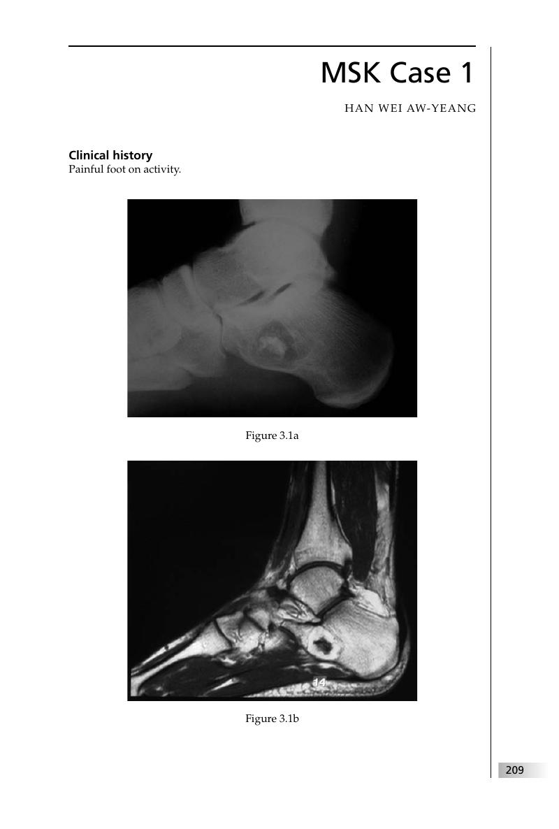
Image of the first page of this content. For PDF version, please use the ‘Save PDF’ preceeding this image.'
Type
Chapter
Information
Final FRCR 2B Viva
A Survival Guide
, pp. 209 - 332
Publisher: Cambridge University Press
Print publication year: 2011

Access options

Get access to the full version of this content by using one of the access options below. (Log in options will check for institutional or personal access. Content may require purchase if you do not have access.)

References

Bertram, C, Popken, F, Rütt, J. Intraosseous lipoma of the calcaneus. Langenbecks Arch Surg 2001; 386: 313–17.CrossRefGoogle ScholarPubMed
Kwak, HS, Lee, KB, Lee, SY, Han, YM. MR findings of calcaneal intraosseous lipoma with hemorrhage. AJR Am J Roentgenol 2005; 185: 1378–9.CrossRefGoogle ScholarPubMed
Murphey, MD, Carroll, JF, Flemming, DJ, et al. From the archives of the AFIP. Benign musculoskeletal lipomatous lesions. Radiographics 2004; 24: 1433–66.CrossRefGoogle ScholarPubMed
Tachdjian, MO. Freiberg's infraction. In Pediatric Orthopedics, 2nd edn. Philadelphia, PA:Saunders, 1990; 1006–10.Google Scholar
Thompson, FM, Hamilton, WG. Problems of the second metatarsophalangeal joint. Orthopedics 1987; 10: 83–9.Google ScholarPubMed
Gordon, L.Paget's disease of the patella. J Bone Joint Surg Am 1958; 40: 1423–5.CrossRefGoogle ScholarPubMed
Smith, SE, Murphey, MD, Motamedi, K, et al. From the archives of the AFIP. Radiologic spectrum of Paget disease of bone and its complications with pathologic correlation. Radiographics 2002; 22: 1191–216.CrossRefGoogle ScholarPubMed
Stull, MA, Moser, RP, Vinh, TN, Kransdorf, MJ, Callaghan, JJ. Paget's disease of the patella. Skeletal Radiol 1990; 19: 407–10.CrossRefGoogle ScholarPubMed
Helms, CA. The meniscus: recent advances in MR imaging of the knee. AJR Am J Roentgenol 2002; 179: 1115–22.CrossRefGoogle Scholar
Ryu, KN, Kim, IS, Kim, EJ. MR imaging of tears of discoid lateral menisci. AJR Am J Roentgenol 1998; 171: 963–7.CrossRefGoogle ScholarPubMed
Gor, DM. Signs in imaging: the trough line sign. Radiology 2002; 224: 485–6.CrossRefGoogle ScholarPubMed
Griffith, JF, Antonio, GE, Yung, PS, et al. Prevalence, pattern, and spectrum of glenoid bone loss in anterior shoulder dislocation: CT analysis of 218 patients. AJR Am J Roentgenol 2008; 190: 1247–54.CrossRefGoogle ScholarPubMed
Saupe, N, White, LM, Bleakney, R, et al. Acute traumatic posterior shoulder dislocation: MR findings. Radiology 2008; 248: 185–93.CrossRefGoogle ScholarPubMed
Blacksin, MF, Finzel, KC, Benevenia, J.Osteomyelitis originating in and around bone infarcts: giant sequestrum phenomena. AJR Am J Roentgenol 2001; 176: 387–91.CrossRefGoogle ScholarPubMed
Hara, H, Akisue, T, Fujimoto, T, et al. Magnetic resonance imaging of medullary bone infarction in the early stage. Clin Imaging 2008; 32: 147–51.CrossRefGoogle ScholarPubMed
Zurlo, JV. The double-line sign. Radiology 1999; 212: 541–2.CrossRefGoogle ScholarPubMed
Balériaux, DL, Neugroschl, C. Spinal and spinal cord infection. Eur Radiol 2004; 14: E72–83Google ScholarPubMed
Conaughty, JM, Chen, J, Martinez, OV, et al. Efficacy of linezolid versus vancomycin in the treatment of methicillin-resistant Staphylococcus aureus discitis: a controlled animal model. Spine 2006; 31: E830–2.CrossRefGoogle ScholarPubMed
Cottle, L, Riordan, T. Infectious spondylodiscitis. J Infect 2008; 56: 401–12.CrossRefGoogle ScholarPubMed
Ehara, S.Spondylodiskitis. AJR Am J Roentgenol 1999; 172: 1450–1.CrossRefGoogle ScholarPubMed
Olsen, KM, Chew, FS. Tumoral calcinosis: pearls, polemics, and alternative possibilities. Radiographics 2006; 26: 871–85.CrossRefGoogle ScholarPubMed
Barron, D, Branfoot, T. Imaging trauma of the appendicular skeleton. Imaging 2003; 15: 324–40.CrossRefGoogle Scholar
Jacobson, JA, Girish, G, Jiang, Y, Sabb, BJ. Radiographic evaluation of arthritis: degenerative joint disease and variations. Radiology 2008; 248; 737–47.CrossRefGoogle ScholarPubMed
Murphey, MD, Carroll, JF, Flemming, DJ, et al. From the archives of the AFIP: benign musculoskeletal lipomatous lesions. Radiographics 2004; 24: 1433–66.CrossRefGoogle ScholarPubMed
Brant, EE, Jordan, HH. Radiologic aspects of hemophilic pseudotumors in bone. Am J Roentgenol Radium Ther Nucl Med 1972; 115: 525–39.Google ScholarPubMed
James, TT, Stafford, JM, Allen, AM, Dixon, LR. Hemophilic pseudotumor: radiologic–pathologic correlation. Radiographics 2003; 23: 852–6.Google Scholar
Park, JS, Ryu, KN. Hemophilic pseudotumor involving the musculoskeletal system: spectrum of radiologic findings. AJR Am J Roentgenol 2004; 183: 55–61.CrossRefGoogle ScholarPubMed
Schreiber, A, Smith, WL, Ionasescu, V, et al. Magnetic resonance imaging of children with Duchenne muscular dystrophy. Pediatric Radiology 1987; 17: 495–7.CrossRefGoogle ScholarPubMed
Chew, FS, Huang-Hellinger, F. Brown tumor. AJR Am J Roentgenol 1993; 160: 752.CrossRefGoogle ScholarPubMed
Eisenberg, RL. Bubbly lesions of bone. AJR Am J Roentgenol 2009; 193: W79–94.CrossRefGoogle Scholar
Miller, TT. Bone tumors and tumorlike conditions: analysis with conventional radiography. Radiology 2008: 246; 662–74.CrossRefGoogle ScholarPubMed
Grainger, AJ, McGonagle, D. Imaging in rheumatology. Imaging 2003; 15: 286–97.CrossRefGoogle Scholar
Jacobson, JA, Girish, G, Jiang, Y, Sabb, BJ. Radiographic evaluation of arthritis: degenerative joint disease and variations. Radiology 2008: 248: 737–47.CrossRefGoogle ScholarPubMed
Sheldon, PJ, Forrester, DM, Learch, TJ. Imaging of intraarticular masses. Radiographics 2005; 25: 105–19.CrossRefGoogle ScholarPubMed
McKenzie, G, Raby, N, Ritchie, D. A pictorial review of primary synovial osteochondromatosis. Eur Radiol 2008; 18: 2662–9.CrossRefGoogle ScholarPubMed
Sheldon, PJ, Forrester, DM, Learch, TJ. Imaging of intraarticular masses. Radiographics 2005; 25: 105–19.CrossRefGoogle ScholarPubMed
Garner, HW, Ortiguera, CJ, Nakhleh, RE. Pigmented villonodular synovitis. Radiographics 2008; 28: 1519–23.Google ScholarPubMed
Murphey, MD, Rhee, JH, Lewis, RB, et al. Pigmented villonodular synovitis: radiologic–pathologic correlation. Radiographics 2008; 28: 1493–518.CrossRefGoogle ScholarPubMed
Sheldon, PJ, Forrester, DM, Learch, TJ. Imaging of intraarticular masses. Radiographics 2005; 25: 105–19.CrossRefGoogle ScholarPubMed
Erlemann, R.Imaging and differential diagnosis of primary bone tumors and tumor-like lesions of the spine. Eur J Radiol 2006; 58: 48–67.CrossRefGoogle ScholarPubMed
Patel, MB, Bhalla, A. Paget's disease of the mandible. N Engl J Med 2008; 358: 625.CrossRefGoogle ScholarPubMed
Leu, KM, Kalemkerian, GP. Osteoporosis circumscripta. N Engl J Med 2002; 347(5): e2.CrossRef
Love, C, Din, AS, Tomas, MB, Kalapparambath, TP, Palestro CJ. Radionuclide bone imaging: an illustrative review. Radiographics 2003; 23: 341–58.CrossRefGoogle Scholar
Smith, SE, Murphey, MD, Motamedi, K, et al. From the archives of the AFIP. Radiologic spectrum of Paget disease of bone and its complications with pathologic correlation. Radiographics 2002; 22: 1191–216.CrossRefGoogle ScholarPubMed
Ejindu, VC, Hine, AL, Mashayekhi, M, Shorvon, PJ, Misra, RR. Musculoskeletal manifestations of sickle cell disease. Radiographics 2007; 27: 1005–21.CrossRefGoogle ScholarPubMed
Lonergan, GJ, Cline, DB, Abbondanzo, SL.Sickle cell anemia. Radiographics 2001; 21: 971–94.CrossRefGoogle ScholarPubMed
Madani, G, Papadopoulou, A, Holloway, B, et al. The radiological manifestations of sickle cell disease. Clin Radiol 2007; 62: 528–38.CrossRefGoogle ScholarPubMed
McCann, C, Mondal, D, Curtis, J. 2_148 The Musculoskeletal Manifestations of the Haemoglobinopathies. Radiology Integrated Training Initiative. Royal College of Radiologists, 2010.
Resnick, D. Hemoglobinopathies and other anemias. In Resnick, D, ed., Diagnosis of Bone and Joint Disorders, 4th edn. Philadelphia, PA: Saunders, 2002; 2146–87.Google Scholar
Angtuaco, EJ, Fassas, AB, Walker, R, Sethi, R, Barlogie, B. Multiple myeloma: clinical review and diagnostic imaging. Radiology 2004; 231: 11–23.CrossRefGoogle ScholarPubMed
Afshani, E, Kuhn, JP. Common causes of low back pain in children. Radiographics 1991; 11: 269–91.CrossRefGoogle ScholarPubMed
Bassett, LW, Blocka, KLN, Furst, , Clements PJ, Gold RH. Skeletal findings in progressive systemic sclerosis (scleroderma). AJR Am J Roentgenol 1981; 136: 1121–6.CrossRefGoogle Scholar
Jones, SN, Stoker, DJ. Radiology at your fingertips: lesions of the terminal phalanx. Clin Radiol 1988; 39: 478–85.CrossRefGoogle ScholarPubMed
Dyer, RB, Chen, MYM, Zagoria, RJ. Classic signs in uroradiology. Radiographics 2004; 24: S247–80.CrossRefGoogle ScholarPubMed
Dyer, RB, Chen, MYM, Zagoria, RJ. Intravenous urography: technique and interpretation. Radiographics 2001; 21: 799–824.CrossRefGoogle ScholarPubMed
Hartman, MS. The golf ball-on-tee sign. Radiology 2006; 239: 297–8.CrossRefGoogle ScholarPubMed
Jung, DC, Kim, SH, Jung, SI, Hwang, SI, Kim, SH. Renal papillary necrosis: review and comparison of findings at multi-detector row CT and intravenous urography. Radiographics 2006; 26: 1827–36.CrossRefGoogle ScholarPubMed
Lonergan, GJ, Cline DB, Abbondanzo SL. Sickle cell anemia. Radiographics 2001; 21: 971–94.CrossRefGoogle ScholarPubMed
Adams, JE. Renal bone disease: radiological investigation. Kidney Int Suppl 1999; 56: S38–41.CrossRefGoogle Scholar
Adams, JE. Radiology of rickets and osteomalacia. In Feldman, D, Glorieux, FH, Pike, W, eds., Vitamin D, 2nd edn. New York, NY: Elsevier, 2005; 967–94.Google Scholar
Pitt, MJ. Rickets and osteomalacia. In Resnick, D, Kransdorf, MJ, eds., Bone and Joint Imaging, 3rd edn. Richmond, VA: Elsevier-Saunders, 2005; 563–75.Google Scholar
Gilula, . Carpal injuries: analytic approach and case exercises. AJR Am J Roentgenol 1979; 133: 503–17.CrossRefGoogle ScholarPubMed
Goldfarb, CA, Yin, Y, Gilula, , Fisher, AJ, Boyer, MI. Wrist fractures: what the clinician wants to know. Radiology 2001; 219: 11–28.CrossRefGoogle Scholar
Kaewlai, R, Avery, LL, Asrani, AV, et al. Multidetector CT of carpal injuries: anatomy, fractures, and fracture-dislocations. Radiographics 2008; 28: 1771–84.CrossRefGoogle ScholarPubMed
Peh, WCG, Galloway, HR. Imaging of wrist injuries. In Vanhoenacker FM, Maas M, Gielen JL, eds., Imaging of Orthopedic Sports Injuries. Berlin: Springer, 2007; 201–24.Google Scholar
Glass, RB, Norton, KI, Mitre, SA, Kang, E. Pediatric ribs: a spectrum of abnormalities. Radiographics 2002; 22: 87–104.CrossRefGoogle ScholarPubMed
Guttentag, AR, Salwen, JK. Keep your eyes on the ribs: the spectrum of normal variants and diseases that involves the ribs. Radiographics 1999; 19: 1125–42.CrossRefGoogle Scholar
Levine, BD, Motamedi, K, Chow, K, Gold, RH, Seeger, LL. CT of rib lesions. AJR Am J Roentgenol 2009; 193: 5–13.CrossRefGoogle ScholarPubMed
David, R, Oria, RA, Kumar, R, et al. Radiologic features of eosinophilic granuloma of bone. AJR Am J Roentgenol 1989; 153: 1021–6.CrossRefGoogle ScholarPubMed
Hoover, KB, Rosenthal, DI, Mankin, H. Langerhans cell histiocytosis. Skeletal Radiol 2007; 36: 95–104CrossRefGoogle ScholarPubMed
Stull, MA, Kransdorf, MJ, Devaney, KO. Langerhans cell histiocytosis of bone. Radiographics 1992; 12: 801–23.CrossRefGoogle Scholar
Burrill, J, Williams, CJ, Bain, G, et al. Tuberculosis: a radiologic review. Radiographics 2007; 27: 1255–73.CrossRefGoogle ScholarPubMed
Engin, G, Acunas, B, Acunas, G, Tunaci, M. Imaging of extrapulmonary tuberculosis. Radiographics 2000; 20: 471–88.CrossRefGoogle ScholarPubMed
Harisinghani, MG, McLoud, TC, Shepard, JA, et al. Tuberculosis from head to toe. Radiographics 2000; 20: 449–70.CrossRefGoogle ScholarPubMed
Jung, NY, Jee, WH, Ha, KY, Park, CK, Byun, JY. Discrimination of tuberculous spondylitis from pyogenic spondylitis on MRI. AJR Am J Roentgenol 2004; 182: 1405–10.CrossRefGoogle ScholarPubMed
Banks, KP, Bui-Mansfield, LT, Chew, FS, Collinson, F. A compartmental approach to the radiographic evaluation of soft-tissue calcifications. Semin Roentgenol 2005; 40: 391–407.CrossRefGoogle ScholarPubMed
Olsen, KM, Chew, FS. Tumoral calcinosis: pearls, polemics, and alternative possibilities. Radiographics 2006; 26: 871–85.CrossRefGoogle ScholarPubMed
Flach, HZ, Ginai, AZ, Oosterhuis, JW. Maffucci syndrome: radiologic and pathologic findings. Radiographics 2001; 21: 1311–16.Google Scholar
Murphey, MD, Flemming, DJ, Boyea, SR, et al. Enchondroma versus chondrosarcoma in the appendicular skeleton: differentiating features. Radiographics 1998; 18: 1213–37.CrossRefGoogle ScholarPubMed
Levine, SM, Lambiase, RE, Petchprapa, CN. Cortical lesions of the tibia: characteristic appearances at conventional radiography. Radiographics 2003; 23: 157–77.CrossRefGoogle ScholarPubMed
Murphey, MD, Choi, JJ, Kransdorf, MJ, Flemming, DJ, Gannon, FH. Imaging of osteochondroma: variants and complications with radiologic-pathologic correlation. Radiographics 2000; 20: 1407–34.CrossRefGoogle ScholarPubMed
Jones, EA, Manaster, BJ, May, DA, Disler, DG. Neuropathic osteoarthropathy: diagnostic dilemmas and differential diagnosis. Radiographics 2000; 20: S279–93.CrossRefGoogle ScholarPubMed
Campos, JC, Chung, CB, Lektrakul, N, et al. Pathogenesis of the Segond fracture: anatomic and MR imaging evidence of an iliotibial tract or anterior oblique band avulsion. Radiology 2001; 219: 381–6.CrossRefGoogle ScholarPubMed
Crotty, JM, Snow, RD, Brogdon, BG, DeMouy, EH. Magnetic resonance imaging of trauma patterns in the knee. Emerg Radiol 1998; 4: 237–44.CrossRefGoogle Scholar
Goldman, AB, Pavlov, H, Rubenstein, D. The Segond fracture of the proximal tibia: a small avulsion that reflects major ligamentous damage. AJR Am J Roentgenol 1988; 151: 1163–7.CrossRefGoogle ScholarPubMed
Matherne, TH, Monu, JUV, Schruff, L, Neitzschman, HR. Avulsions around the knee portend instability. Emerg Radiol 2005; 11: 213–18.CrossRefGoogle ScholarPubMed
Miller, LS, Yu, JS. Radiographic indicators of acute ligament injuries of the knee: a mechanistic approach. Emerg Radiol 2010; 17: 435–44.CrossRefGoogle ScholarPubMed
Miller, TT. Bone tumors and tumorlike conditions: analysis with conventional radiography. Radiology 2008; 246: 662–74.CrossRefGoogle ScholarPubMed
Resnick, D. Diagnosis of Bone and Joint Disorders, 4th edn. Philadelphia, PA: Saunders, 2002; 3757, 3922–4.Google Scholar
Chew, FS, Roberts CC, Lalaji AP. Musculoskeletal Imaging: a Teaching File, 2nd edn. Philadelphia, PA:Lippincott Williams & Wilkins, 2006.Google Scholar
Helliwell, PS, Taylor, WJ. Classification and diagnostic criteria for psoriatic arthritis. Ann Rheum Dis 2005; 64: ii3–8.CrossRefGoogle ScholarPubMed
Ory, PA, Gladman DD, Mease PJ. Psoriatic arthritis and imaging. Ann Rheum Dis 2005; 64: ii55–7CrossRefGoogle Scholar
Chew, FS, Huang-Hellinger, F. Brown tumor. AJR Am J Roentgenol 1993; 160: 752.CrossRefGoogle ScholarPubMed
Eisenberg, RL. Bubbly lesions of bone. AJR Am J Roentgenol 2009; 193: W79–94.CrossRefGoogle Scholar
Jetvic, V. Imaging of renal osteodystrophy. Eur J Radiol 2003; 46: 85–95.Google Scholar
Jones, SN, Stoker, DJ. Radiology at your fingertips: lesions of the terminal phalanx. Clin Radiol 1988; 39: 478–85.CrossRefGoogle ScholarPubMed
McDonald, DK, Parman, L, Speights, VO. Best cases from the AFIP: primary hyperparathyroidism due to parathyroid adenoma. Radiographics 2005; 25: 829–34.CrossRefGoogle ScholarPubMed
Miller, TT. Bone tumors and tumorlike conditions: analysis with conventional radiography. Radiology 2008; 246: 662–74.CrossRefGoogle ScholarPubMed
Tigges, S, Nance, EP, Carpenter, WA, Erb, R. Osteodystrophy: imaging findings that mimic those of other diseases,AJR Am J Roentgenol 1995; 165: 143–8.CrossRefGoogle Scholar
Wittenberg, A. The rugger jersey spine sign. Radiology 2004; 230: 491–2.CrossRefGoogle ScholarPubMed
Chew, FS. Radiology of the hands: review and self-assessment module. AJR Am J Roentgenol 2005; 184: S157–68.CrossRefGoogle ScholarPubMed
Koyama, T, Ueda, H, Togashi, K, et al. Radiologic manifestations of sarcoidosis in various organs. Radiographics 2004; 24: 87–104.CrossRefGoogle ScholarPubMed
Moore, SL, Teirstein, AE. Musculoskeletal sarcoidosis: spectrum of appearances at mr imaging. Radiographics 2003; 23: 1389–99.CrossRefGoogle ScholarPubMed
Resnick, D, Niwayama, G. Sarcoidosis. In Resnick, D, Niwayama, G, eds., Diagnosis of Bone and Joint Disorders, 3rd edn. Philadelphia, PA: Saunders, 1995; 4333–52.Google Scholar
Yaghmai, I. Radiographic, angiographic and radionuclide manifestations of osseous sarcoidosis. Radiographics 1983; 3: 375–96.CrossRefGoogle Scholar
Littrell, , Wenger, , Wold, , et al. Radiographic, CT, and MR imaging features of dedifferentiated chondrosarcomas: a retrospective review of 174 de novo cases. Radiographics 2004; 24: 1397–409.CrossRefGoogle Scholar
Murphey, MD, Walker, EA, Wilson, AJ, et al. From the archives of the AFIP. Imaging of primary chondrosarcoma: radiologic–pathologic correlation. Radiographics 2003; 23: 1245–78.CrossRefGoogle ScholarPubMed
Eisenberg, RL. Bubbly lesions of bone. AJR Am J Roentgenol 2009; 193: W79–94.CrossRefGoogle Scholar
Miller, TT. Bone tumors and tumorlike conditions: analysis with conventional radiography. Radiology 2008; 246: 662–74.CrossRefGoogle ScholarPubMed
Murphey, MD, Nomikos, GC, Flemming, DJ, et al. From the archives of the AFIP. Imaging of giant cell tumor and giant cell reparative granuloma of bone: radiologic– pathologic correlation. Radiographics 2001; 21: 1283–309.CrossRefGoogle ScholarPubMed
Ashlock, SJ, Harris, JH. West, OC, Zelitt, D. Acute elbow injuries. Emerg Radiol 1998; 5: 416–37.CrossRefGoogle Scholar
Barron, D, Branfoot, T. Imaging trauma of the appendicular skeleton. Imaging 2003; 15: 324–40.CrossRefGoogle Scholar
Gleeson, AP, Beattie, TF. Monteggia fracture–dislocation in children. J Accid Emerg Med 1994; 11: 192–4.CrossRefGoogle ScholarPubMed
Hunter, TB, Peltier, LF, Lund, PJ.Radiologic history exhibit. Musculoskeletal eponyms: who are those guys?Radiographics 2000; 20: 819–36.CrossRefGoogle ScholarPubMed
John, SD, Wherry, K, Swischuk, Phillips WA. Improving detection of pediatric elbow fractures by understanding their mechanics. Radiographics 1996; 16: 1443–60.CrossRefGoogle ScholarPubMed
Camp, MD, Tompkins, RK, Spanier, SS, Bridge, JA, Bush, CH. Best cases from the AFIP: adamantinoma of the tibia and fibula with cytogenetic analysis. Radiographics 2008; 28: 1215–20.Google ScholarPubMed
Eisenberg, RL. Bubbly lesions of bone. AJR Am J Roentgenol 2009; 193: W79–94.CrossRefGoogle Scholar
Levine, SM, Lambiase RE, Petchprapa CN.Cortical lesions of the tibia: characteristic appearances at conventional radiography. Radiographics 2003; 23: 157–77.CrossRefGoogle ScholarPubMed
Miller, TT. Bone tumors and tumorlike conditions: analysis with conventional radiography. Radiology 2008; 246: 862–74.CrossRefGoogle ScholarPubMed
O'Donnell, P.Evaluation of focal bone lesions: basic principles and clinical scenarios. Imaging 2003; 15: 298–323.CrossRefGoogle Scholar
Chai, JW, Hong, SH, Choi, JY, et al. Radiologic diagnosis of osteoid osteoma: from simple to challenging findings. Radiographics 2010; 30: 737–49.CrossRefGoogle ScholarPubMed
Kransdorf, MJ, Stull, MA, Gilkey, FW, Moser, RP. From the archives of the AFIP: osteoid osteoma. Radiographics 1991; 11: 671–96.CrossRefGoogle Scholar
Ashlock, SJ, Harris, JH, West OC, Zelitt D. Acute elbow injuries. Emerg Radiol 1998; 5: 416–37.CrossRefGoogle Scholar
John, SD, Wherry, K, Swischuk, Phillips WA. Improving detection of pediatric elbow fractures by understanding their mechanics. Radiographics 1996; 16: 1443–60.CrossRefGoogle ScholarPubMed
Dunfee, BL, Sakai, O, Pistey, R, Gohel, A. Radiologic and pathologic characteristics of benign and malignant lesions of the mandible. Radiographics 2006; 26: 1751–68.CrossRefGoogle ScholarPubMed
Murphey, MD, Robbin, MR, McRae, GA, et al. The many faces of osteosarcoma. Radiographics 1997; 17: 1205–31.Google ScholarPubMed
Rana, RS, Wu, JS, Eisenberg, RL. Pattern of the month: periosteal reaction. AJR Am J Roentgenol 2009; 193: W259–72.CrossRefGoogle Scholar
Lin, J, Martel, W. Cross-sectional imaging of peripheral nerve sheath tumors: characteristic signs on CT, MR imaging, and sonography. AJR Am J Roentgenol 2001; 176: 75–82.CrossRefGoogle ScholarPubMed
Tateishi, U, Gladish, GW, Kusumoto, M, et al. Chest wall tumors: radiologic findings and pathologic correlation. Part 1. Benign tumors. Radiographics 2003; 23: 1477–90.CrossRefGoogle ScholarPubMed

Save book to Kindle

To save this book to your Kindle, first ensure [email protected] is added to your Approved Personal Document E-mail List under your Personal Document Settings on the Manage Your Content and Devices page of your Amazon account. Then enter the ‘name’ part of your Kindle email address below. Find out more about saving to your Kindle.

Note you can select to save to either the @free.kindle.com or @kindle.com variations. ‘@free.kindle.com’ emails are free but can only be saved to your device when it is connected to wi-fi. ‘@kindle.com’ emails can be delivered even when you are not connected to wi-fi, but note that service fees apply.

Find out more about the Kindle Personal Document Service.

Available formats
×

Save book to Dropbox

To save content items to your account, please confirm that you agree to abide by our usage policies. If this is the first time you use this feature, you will be asked to authorise Cambridge Core to connect with your account. Find out more about saving content to Dropbox.

Available formats
×

Save book to Google Drive

To save content items to your account, please confirm that you agree to abide by our usage policies. If this is the first time you use this feature, you will be asked to authorise Cambridge Core to connect with your account. Find out more about saving content to Google Drive.

Available formats
×