Skip to main content Accessibility help
×
Hostname: page-component-78c5997874-t5tsf Total loading time: 0 Render date: 2024-11-10T05:37:02.146Z Has data issue: false hasContentIssue false

2 - Gastrointestinal radiology

Published online by Cambridge University Press:  05 March 2012

Kiat Tsong Tan
Affiliation:
Queen Elizabeth Hospital, Charlottetown
John Curtis
Affiliation:
University Hospital Aintree
Jessie Aw
Affiliation:
University of Chicago
Get access

Summary

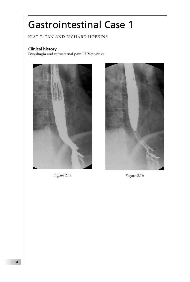
Image of the first page of this content. For PDF version, please use the ‘Save PDF’ preceeding this image.'
Type
Chapter
Information
Final FRCR 2B Viva
A Survival Guide
, pp. 114 - 208
Publisher: Cambridge University Press
Print publication year: 2011

Access options

Get access to the full version of this content by using one of the access options below. (Log in options will check for institutional or personal access. Content may require purchase if you do not have access.)

References

Federle, M, Jeffrey, RB, Anne, VB. Diagnostic Imaging of the Abdomen. Salt Lake City, UT: Amirsys, 2004.Google Scholar
Roberts, L, Gibbons, R, Gibbons, G, Rice, RP, Thompson, WM. Adult esophageal candidiasis: a radiographic spectrum. Radiographics 1987; 7: 289–307.CrossRefGoogle ScholarPubMed
Levine, MS, Rubesin SE. Diseases of the esophagus: diagnosis with esophagography. Radiology 2005; 237: 414–27.CrossRefGoogle ScholarPubMed
Woodfield, CA, Levine, MS, Rubesin, SE, Langlotz, CP, Laufer, I. Diagnosis of primary versus secondary achalasia: reassessment of clinical and radiographic criteria. AJR Am J Roentgenol 2000; 175: 727–31.CrossRefGoogle ScholarPubMed
Levine, MS, Moolten, DN, Herlinger, H, Laufer, I. Esophageal intramural pseudodiverticulosis: a reevaluation. AJR Am J Roentgenol 1986; 147: 1165–70.CrossRefGoogle Scholar
Mahajan, SK, Warshauer, DM, Bozymski, EM. Esophageal intramural pseudo-diverticulosis: endoscopic and radiologic correlation. Gastrointest Endosc 1993; 39: 565–7.CrossRefGoogle ScholarPubMed
White, CS, Templeton, PA, Attar, S. Esophageal perforation: CT findings. AJR Am J Roentgenol 1993; 160: 767–70.CrossRefGoogle ScholarPubMed
Young, CA, Menias, CO, Bhalla, S, Prasad, SR. CT features of esophageal emergencies. Radiographics 2008; 28: 1541–53.CrossRefGoogle ScholarPubMed
Woodring, JH. Unusual manifestations of lung cancer. Radiol Clin North Am 1990; 28: 599–618.
Gupta, AA, Kim, DC, Krinsky, GA, Lee, VS. CT and MRI of cirrhosis and its mimics. AJR Am J Roentgenol 2004; 183: 1595–601.CrossRefGoogle ScholarPubMed
Kawamoto, S, Soyer, PA, Fishman, EK, Bluemke, DA. Nonneoplastic liver disease: evaluation with CT and MR imaging. Radiographics 1998; 18: 827–48.CrossRefGoogle ScholarPubMed
Hewitt, AN, Levine, MS, Rubesin, SE, Laufer, I. Gastric bezoars: reassessment of clinical and radiographic findings in 19 patients. Br J Radiol 2009; 82: 901–7.CrossRefGoogle ScholarPubMed
Ghai, S, Pattison, J, Ghai, S, et al. Primary gastrointestinal lymphoma: spectrum of imaging findings with pathologic correlation. Radiographics 2007; 27: 1371–88.CrossRefGoogle ScholarPubMed
Levine, MS. Benign tumors. In: Gore, RM, Levine, MS, Laufer, I, eds., Textbook of Gastrointestinal Radiology. Philadelphia, PA: Saunders, 1994; 628–58.Google Scholar
Wagner, BJ, Brower, AC. General case of the day: gastric hyperplastic polyps. Radiographics 1994; 14: 682–5.CrossRefGoogle ScholarPubMed
Levy, AD, Remotti, HE, Thompson, WM, Sobin, LH, Miettinen, M.Gastrointestinal stromal tumors: radiologic features with pathologic correlation. Radiographics 2003; 23: 283–304.CrossRefGoogle ScholarPubMed
Sandrasegaran, K, Rajesh, A, Rydberg, J, et al. Gastrointestinal stromal tumors: clinical, radiologic, and pathologic features. AJR Am J Roentgenol 2005; 184: 803–11.CrossRefGoogle ScholarPubMed
Mortelé, KJ, Rocha, TC, Streeter, JL, Taylor, AJ. Multimodality imaging of pancreatic and biliary congenital anomalies. Radiographics 2006; 26: 715–31.CrossRefGoogle ScholarPubMed
Gallego, C, Velasco, M, Marcuello, P, et al. Congenital and acquired anomalies of the portal venous system. Radiographics 2002; 22: 141–59.CrossRefGoogle ScholarPubMed
Ito, K, Higuchi, M, Kada, T, et al. CT of acquired abnormalities of the portal venous system. Radiographics 1997; 17: 897–917.CrossRefGoogle ScholarPubMed
Choi, BG, Park, SH, Byun, JY, et al. The findings of ruptured hepatocellular carcinoma on helical CT. Br J Radiol 2001; 74: 142–6.CrossRefGoogle ScholarPubMed
Orwig, D, Federle, MP. Localized clotted blood as evidence of visceral trauma on CT: the sentinel clot sign. AJR Am J Roentgenol 1989; 153: 747–9.CrossRefGoogle ScholarPubMed
Hussain, SM, Terkivatan, T, Zondervan, PE, et al. Focal nodular hyperplasia: findings at state-of-the-art MR imaging, US, CT, and pathologic analysis. Radiographics 2004; 24: 3–17.CrossRefGoogle ScholarPubMed
Kamel, IR, Liapi, E, Fishman, EK. Focal nodular hyperplasia: lesion evaluation using 16-MDCT and 3D CT angiography. AJR Am J Roentgenol 2006; 186: 1587–96.CrossRefGoogle ScholarPubMed
Summerton, SL, Hollander, AC, Stassi, J, Rosenberg, HK, Carroll, SF. US case of the day: gallstone ileus. Radiographics 1995; 15: 493–5.CrossRefGoogle ScholarPubMed
Gibson, RN. The biliary system. In Adam, A, Dixon, AK, eds., Grainger and Allison's Diagnostic Radiology, 5th edn. London: Churchill Livingstone, 2008 (ebook).Google Scholar
Irie, H, Honda, H, Kuroiwa, T, et al. Pitfalls in MR cholangiopancreatographic interpretation. Radiographics 2001; 21: 23–37.CrossRefGoogle ScholarPubMed
Nandalur, KR, Hussain, HK, Weadock, WJ, et al. Possible biliary disease: diagnostic performance of high-spatial-resolution isotropic 3D T2-weighted MRCP. Radiology 2008; 249: 883–90.CrossRefGoogle ScholarPubMed
Grand, D, Horton, KM, Fishman, E. CT of the gallbladder: spectrum of disease. AJR Am J Roentgenol 2004; 183: 163–70.CrossRefGoogle Scholar
Stephen, AE, Berger, DL. Carcinoma in the porcelain gallbladder: a relationship revisited. Surgery 2001; 129: 699–703.CrossRefGoogle ScholarPubMed
Grand, D, Horton, KM, Fishman, E. CT of the gallbladder: spectrum of disease. AJR Am J Roentgenol 2004; 183: 163–70.CrossRefGoogle Scholar
Levy, AD, Murakata, , Rohrmann, CA. Gallbladder carcinoma: radiologic–pathologic correlation. Radiographics 2001; 21: 295–314.CrossRefGoogle ScholarPubMed
Misra, S, Chaturvedi, A, Misra, NC, Sharma, ID. Carcinoma of the gallbladder. Lancet Oncol 2003; 4: 167–76.CrossRefGoogle ScholarPubMed
Cho, GJ, Bergquist, K, Schwartz, AM. Peutz–Jeghers syndrome and hamartomatous polyposis syndrome: radiologic–pathologic correlation. Radiographics 1997; 17: 785–91.CrossRefGoogle ScholarPubMed
Half, E, Bercovich, D, Rozen, P. Familial adenomatous polyposis. Orphanet J Rare Dis 2009; 4: 22.CrossRefGoogle ScholarPubMed
Gore, RM, Balthazar, EJ, Ghahremani, CG, Miller, FH. CT features of ulcerative colitis and Crohn's disease. AJR Am J Roentgenol 1996; 167: 3–15.CrossRefGoogle ScholarPubMed
Halligan, S. The small bowel and peritoneal cavity. In Sutton, D, ed., Textbook of Radiology and Imaging, 7th edn. London: Elsevier, 2003; 615–34.Google Scholar
Buckland, JA, Fishman, EK. CT evaluation of small bowel neoplasms: spectrum of disease. Radiographics 1999; 18: 379–92.Google Scholar
Lee, WK, Lau, EW, Duddalwar, VA, Stanley, AJ, Ho, YY. Abdominal manifestations of extranodal lymphoma: spectrum of imaging findings. AJR Am J Roentgenol 2008; 191: 198–206.CrossRefGoogle ScholarPubMed
Aguirre, DA, Santosa, AC, Casola, G, Sirlin, CB. Abdominal wall hernias: imaging features, complications, and diagnostic pitfalls at multidetector row CT. Radiographics 2005; 25: 1501–20.CrossRefGoogle Scholar
Mantoo, SK, Mak, K, Tan, TJ. Obturator hernia: diagnosis and treatment in the modern era. Singapore Med J 2009; 50: 866–70.Google ScholarPubMed
Levy, AD, Sobin, LH. From the archives of the AFIP. Gastrointestinal carcinoids: imaging features with clinicopathologic comparison. Radiographics 2007; 27: 237–57.CrossRefGoogle ScholarPubMed
Buetow, PC, Miller, DL, Parrino, TV, Buck, JL. Islet cell tumors of the pancreas: clinical, radiologic, and pathologic correlation in diagnosis and localization. Radiographics 1997; 17: 453–72.CrossRefGoogle ScholarPubMed
Fidler, JL, Fletcher, JG, Reading, CC, et al. Preoperative detection of pancreatic insulinomas on multiphasic helical CT. AJR Am J Roentgenol 2003; 181: 775–80.CrossRefGoogle ScholarPubMed
Lewis, RB, Lattin, GE, Paal, E. Pancreatic endocrine tumors: radiologic-clinicopathologic correlation. Radiographics 2010; 30: 1445–64.CrossRefGoogle ScholarPubMed
Acar, M, Tatli, S. Cystic tumors of the pancreas: a radiological perspective. Diagn Interv Radiol 2011; 17: 143–9.Google ScholarPubMed
Kim, YH, Saini, S, Sahani, D, et al. Imaging diagnosis of cystic pancreatic lesions: pseudocyst versus nonpseudocyst. Radiographics 2005; 25: 671–85.CrossRefGoogle ScholarPubMed
Sahani, DV, Kadavigere, R, Saokar, A, et al. Cystic pancreatic lesions: a simple imaging-based classification system for guiding management. Radiographics 2005; 25: 1471–84.CrossRefGoogle ScholarPubMed
Baillie, J, Paulson, E, Vitellas, K. Biliary imaging: a review. Gastroenterology 2003; 124: 1686–99.CrossRefGoogle ScholarPubMed
Brennan, DD, Zamboni, GA, Raptopoulos, VD, Kruskal, JB. Comprehensive preoperative assessment of pancreatic adenocarcinoma with 64-section volumetric CT. Radiographics 2007; 27: 1653–66.CrossRefGoogle ScholarPubMed
Kim, JH, Kim, MJ, Chung, JJ, et al. Differential diagnosis of periampullary carcinomas at MR imaging. Radiographics 2002; 22: 1335–52.CrossRefGoogle ScholarPubMed
Tamm, EP, Silverman, PM, Charnsangavej, C, Evans, DB. Diagnosis, staging and surveillance of pancreatic cancer. AJR Am J Roentgenol 2003; 180: 1311–23.CrossRefGoogle ScholarPubMed
Demos, TC, Posniak, HV, Harmath, C, Olson, MC, Aranha, G. Cystic lesions of the pancreas. AJR Am J Roentgenol 2002; 179: 1375–88.CrossRefGoogle ScholarPubMed
Sunnapwar, A, Prasad, SR, Menias, CO, et al. Nonalcoholic, nonbiliary pancreatitis: cross-sectional imaging spectrum. AJR Am J Roentgenol 2010; 195: 67–75.CrossRefGoogle ScholarPubMed
Choi, SH, Han, JK, Kim, SH, et al. Intussusception in adults: from stomach to rectum. AJR Am J Roentgenol 2004; 183: 691–8.CrossRefGoogle ScholarPubMed
Kim, YH, Blake, MA, Harisinghani, MG, et al. Adult intestinal intussusceptions: CT appearances and identification of a causative lead point. Radiographics 2006; 26: 733–44.CrossRefGoogle ScholarPubMed
Birnbaum, BA, Jeffrey, RB. CT and sonographic evaluation of acute right lower quadrant abdominal pain. AJR Am J Roentgenol 1998; 170: 361–71.CrossRefGoogle ScholarPubMed
Pinto Leite, N, Pereira, JM, Cunha, R, Pinto, P, Sirlin, C. CT evaluation of appendicitis and its complications: imaging techniques and key diagnostic features. AJR Am J Roentgenol 2005; 185: 406–17.CrossRefGoogle Scholar
Yu, J, Fulcher, AS, Turner, MA, Halvorsen, RA. Helical CT evaluation of acute right lower quadrant pain: part I, common mimics of appendicitis. AJR Am J Roentgenol 2005; 184: 1136–42.CrossRefGoogle ScholarPubMed
Yu, J, Fulcher, AS, Turner, MA, Halvorsen, RA. Helical, CTevaluation of acute right lower quadrant pain: part II, uncommon mimics of appendicitisAJR Am J Roentgenol 2005; 184: 1143–9.CrossRefGoogle ScholarPubMed
Buetow, PC, Buck, JL, Carr, NJ, Pantongrag-Brown, L. From the archives of the AFIP. Colorectal adenocarcinoma: radiologic–pathologic correlation. Radiographics 1995; 15: 127–46.CrossRefGoogle ScholarPubMed
Pickhardt, PJ, Kim, DH. Colorectal cancer screening with CT colonography: key concepts regarding polyp prevalence, size, histology, morphology, and natural history. AJR Am J Roentgenol 2009; 193: 40–6.CrossRefGoogle ScholarPubMed
Silva, AC, Hara, AK, Leighton, JA, Heppell, JP. CT colonography with intravenous contrast material: varied appearances of colorectal carcinoma. Radiographics 2005; 25: 1321–34.CrossRefGoogle ScholarPubMed
Dann, PH, Krinsky, GA, Israel, GM. Case 135: presacral myelolipoma. Radiology 2008; 248: 314–16.CrossRefGoogle ScholarPubMed
Kocaoglu, M, Frush, DP. Pediatric presacral masses. Radiographics 2006; 26: 833–57.CrossRefGoogle ScholarPubMed
Pereira, JM, Sirlin, CB, Pinto, PS, Casola, G. CT and MR imaging of extrahepatic fatty masses of the abdomen and pelvis: techniques, diagnosis, differential diagnosis, and pitfalls. Radiographics 2005; 25: 69–85.CrossRefGoogle Scholar
Harisinghani, MG, Gervais, DA, Hahn, PF, et al. CT-guided transgluteal drainage of deep pelvic abscesses: indications, technique, procedure-related complications, and clinical outcome. Radiographics 2002; 22: 1353–67.CrossRefGoogle ScholarPubMed
Horton, KM, Corl, FM, Fishman, EK. CT evaluation of the colon: inflammatory disease. Radiographics 2000; 20: 399–418.CrossRefGoogle ScholarPubMed
Berrocal, T, Lamas, M, Gutierrez, J, et al. Congenital anomalies of the small intestine, colon and rectum. Radiographics 1999; 19: 1219–36.CrossRefGoogle ScholarPubMed
Long, FR, Kramer, SS, Markowitz, RI, Taylor, GE. Radiographics patterns of intestinal malrotation in children. Radiographics 1996; 16: 547–56.CrossRefGoogle ScholarPubMed
Ambrosini, R, Barchiesi, A, Di Mizio, V, et al. Inflammatory chronic disease of the colon: how to image. Eur J Radiol 2007; 61: 442–8.CrossRefGoogle Scholar
Bartram, CI, Taylor, S. The large bowel. In Adam, A, Dixon, AK, eds., Grainger and Allison's Diagnostic Radiology, 5th edn. London: Churchill Livingstone, 2008 (ebook).Google Scholar
Kawamoto, S, Horton, KM, Fishman, EK. Pseudomembranous colitis: spectrum of imaging findings with clinical and pathologic correlation. Radiographics 1999; 19: 887–97.CrossRefGoogle ScholarPubMed
Balthazar, EJ, Siegel, SE, Megibow, AJ, Scholes, J, Gordon, R. CT in patients with scirrhous carcinoma of the GI tract: imaging findings and value for tumor detection and staging. AJR Am J Roentgenol 1995; 165: 839–45.CrossRefGoogle ScholarPubMed
Halligan, S. The large bowel. In Sutton, D, ed., Textbook of Radiology and Imaging, 7th edn. London: Elsevier, 2003; 635–63.Google Scholar
Consorti, ET, Liu, TH. Diagnosis and treatment of caecal volvulus. Postgrad Med J 2005; 81: 772–6.CrossRefGoogle ScholarPubMed
Perret, RS, Kunberger, . Case 44: cecal volvulus. AJR Am J Roentgenol 1998; 171: 855, 859, 860.CrossRefGoogle Scholar
Peterson, CM, Anderson, JS, Hara, AK, Carenza, JW, Menias, CO.Volvulus of the gastrointestinal tract: appearances at multimodality imaging. Radiographics 2009; 29: 1281–93.CrossRefGoogle ScholarPubMed
Rha, SE, Ha, HK, Lee, SH, et al. CT and MR imaging findings of bowel ischemia from various primary causes. Radiographics 2000; 20: 29–42.CrossRefGoogle Scholar
Sebastia, C, Quiroga, S, Espin, E, et al. Portomesenteric vein gas: pathologic mechanisms, CT findings, and prognosis. Radiographics 2000; 20: 1213–24.CrossRefGoogle ScholarPubMed
Horton, KM, Lawler, LP, Fishman, EK. CT findings in sclerosing mesenteritis (panniculitis): spectrum of disease. Radiographcis 2003; 23: 1561–7.CrossRefGoogle Scholar
Nishino, M, Hayakawa, K, Minami, M, et al. Primary retroperitoneal neoplasms: CT and MR imaging findings with anatomic and pathologic diagnostic clues. Radiographics 2003; 23: 45–57.CrossRefGoogle Scholar
Pereira, JM, Sirlin, CB, Pinto, PS, Casola, G. CT and MR imaging of extrahepatic fatty masses of the abdomen and pelvis: techniques, diagnosis, differential diagnosis, and pitfalls. Radiographics 2005; 25: 69–85.CrossRefGoogle Scholar
Cronin, CG, Lohan, DG, Blake, MA, et al. Retroperitoneal fibrosis: a review of clinical features and imaging findings. AJR Am J Roentgenol 2008; 191: 423–31CrossRefGoogle ScholarPubMed
Georgiades, CS, Neyman, EG, Barish, MA, Fishman, EK. Amyloidosis: review and CT manifestations. Radiographics 2004; 24: 405–16.CrossRefGoogle ScholarPubMed
Kim, SH, Han, JK, Lee, KH, et al. Abdominal amyloidosis: spectrum of radiologic findings. Clin Radiol 2003; 58: 610–20.CrossRefGoogle Scholar
Abdalian, R, Heathcote, EJ. Sclerosing cholangitis: a focus on secondary causes. Hepatology 2006; 44: 1063–74.CrossRefGoogle ScholarPubMed
Vitellas, KM, Keogan, MT, Freed, KS, et al. Radiologic manifestations of sclerosing cholangitis with emphasis on MR cholangiopancreatography 1. Radiographics 2000; 20: 959–75.CrossRefGoogle Scholar
Lubner, M, Menias, C, Rucker, C, et al. Blood in the belly: CT findings of hemoperitoneum. Radiographics 2007; 27: 109–25.CrossRefGoogle ScholarPubMed
Raikhlin, A, Baerlocher, MO, Asch, MR, Myers, A. Imaging and transcatheter arterial embolization for traumatic splenic injuries: review of the literature. Can J Surg 2008; 51: 464–72.Google ScholarPubMed

Save book to Kindle

To save this book to your Kindle, first ensure [email protected] is added to your Approved Personal Document E-mail List under your Personal Document Settings on the Manage Your Content and Devices page of your Amazon account. Then enter the ‘name’ part of your Kindle email address below. Find out more about saving to your Kindle.

Note you can select to save to either the @free.kindle.com or @kindle.com variations. ‘@free.kindle.com’ emails are free but can only be saved to your device when it is connected to wi-fi. ‘@kindle.com’ emails can be delivered even when you are not connected to wi-fi, but note that service fees apply.

Find out more about the Kindle Personal Document Service.

Available formats
×

Save book to Dropbox

To save content items to your account, please confirm that you agree to abide by our usage policies. If this is the first time you use this feature, you will be asked to authorise Cambridge Core to connect with your account. Find out more about saving content to Dropbox.

Available formats
×

Save book to Google Drive

To save content items to your account, please confirm that you agree to abide by our usage policies. If this is the first time you use this feature, you will be asked to authorise Cambridge Core to connect with your account. Find out more about saving content to Google Drive.

Available formats
×