No CrossRef data available.
Article contents
Important steps in a Correlative Light Electron Microscopy Experiment
Published online by Cambridge University Press: 23 September 2015
Abstract
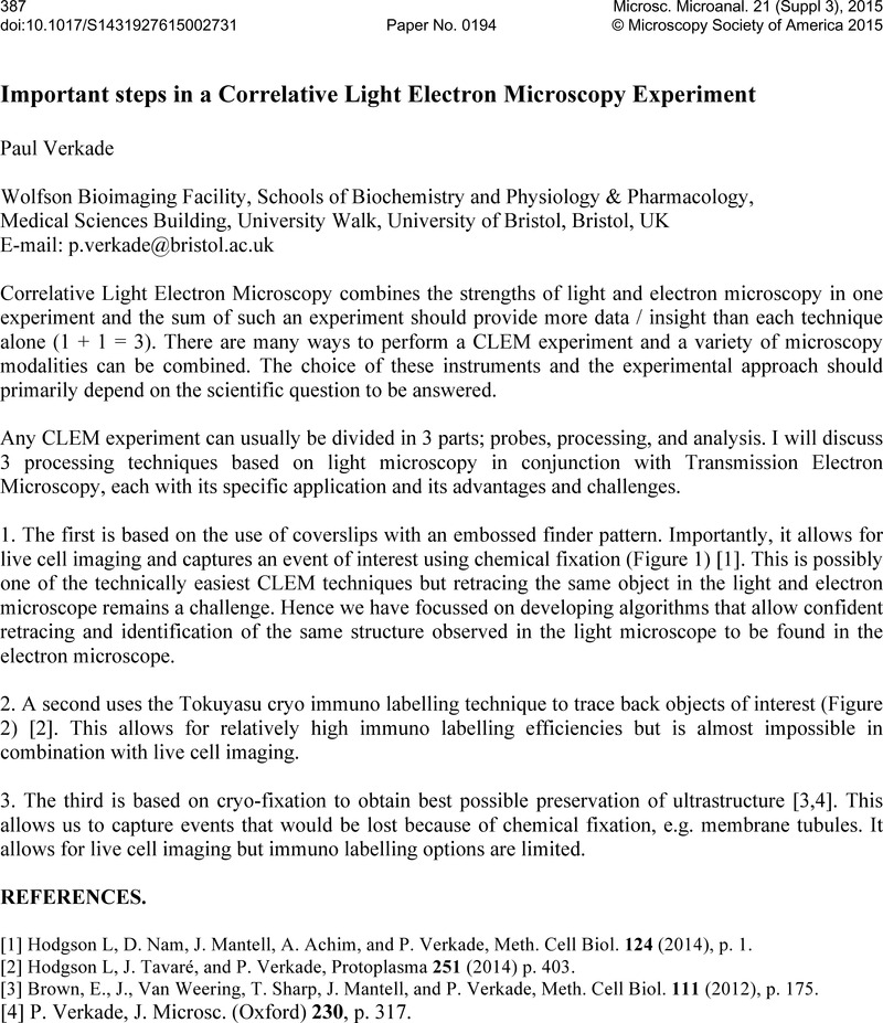
An abstract is not available for this content so a preview has been provided. As you have access to this content, a full PDF is available via the ‘Save PDF’ action button.

- Type
- Abstract
- Information
- Microscopy and Microanalysis , Volume 21 , Supplement S3: Proceedings of Microscopy & Microanalysis 2015 , August 2015 , pp. 387 - 388
- Copyright
- Copyright © Microscopy Society of America 2015
References
REFERENCES
[1]
Hodgson, L, Nam, D., Mantell, J., Achim, A. & Verkade, P., Meth. Cell Biol.
124 (2014). p. 1.Google Scholar
[3]
Brown, E., Van Weering, J., Sharp, T., Mantell, J. & Verkade, P., Meth. Cell Biol.
111 (2012). p. 175.Google Scholar


