Hostname: page-component-586b7cd67f-dlnhk Total loading time: 0 Render date: 2024-11-29T07:58:15.841Z Has data issue: false hasContentIssue false

Systematic Investigation of Lanthanoid Transition Heavy Metal Acetates as Electron Staining Reagents for Protein Molecules in Biological Transmission Electron Microscopy

Published online by Cambridge University Press:  01 April 2022

Noriyuki Ishii*
Affiliation:
Cellular and Molecular Biotechnology Research Institute, Department of Life Science and Biotechnology, National Institute of Advanced Industrial Science and Technology (AIST), Tsukuba Central-6, 1-1-1 Higashi, Tsukuba, Ibaraki 305-8566, Japan Electron Microscopy Facility, Open Research Facilities Station, Open Research Platform Unit, Tsukuba Innovation Arena (TIA) Central Office, National Institute of Advanced Industrial Science and Technology (AIST), 1-1-1 Higashi, Tsukuba, Ibaraki 305-8566, Japan The United Graduate School of Agricultural Science, Gifu University, 1-1 Yanagido, Gifu City, Gifu 501-1193, Japan Human Resources Bureau, National Personnel Authority, 1-2-3 Kasumigaseki, Chiyoda-ku, Tokyo 100-8913, Japan
*
*Corresponding author: Noriyuki Ishii, E-mail: [email protected]
Get access

Abstract

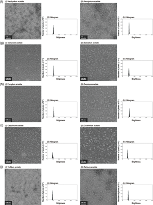
Cryo-electron microscopy, widely used for high-resolution protein structure determination, does not require staining. Yet negative staining with heavy metal salts such as uranyl acetate has been in persistent demand since the 1950s due to its image contrasting capabilities at room temperature with a common electron microscope. However, uranium compounds are nuclear fuel materials and are tightly controlled worldwide. Acetates of each lanthanoid series elements except promethium are prepared at the same concentration (2%(w/v)) and used as a model on horse spleen ferritin for electron microscopic analysis to systematically evaluate their effectiveness as electron staining reagents for the protein. Analysis shows that the triacetates of samarium and europium, followed by gadolinium and erbium, and then lanthanum and neodymium could function as electron staining reagents. Thulium-triacetate precipitates thin plate-like crystals and may be used for selecting better imaging fields. Of the 14 lanthanoid-triacetates examined, about half are viable alternatives to uranyl acetate as an electron staining reagent for ferritin, and there appears an optimal range in ionic sizes for promising lanthanoids. This is the first systematic investigation of lanthanoid transition heavy metal triacetates from the viewpoint of lanthanoid contraction, using density distribution histograms of electron micrographs as an indicator for comparison with uranyl acetate.

Type
Biological Applications
Copyright
Copyright © The Author(s), 2022. Published by Cambridge University Press on behalf of the Microscopy Society of America

Access options

Get access to the full version of this content by using one of the access options below. (Log in options will check for institutional or personal access. Content may require purchase if you do not have access.)

References

Baschong, W & Aebi, U (2006). Negative staining. In Cell Biology: A Laboratory Handbook, 3rd ed, vol. 3, Celis, JE (Ed.), pp. 233240. Cambridge, Massachusetts: Academic Press.CrossRefGoogle Scholar
Biswas, S, Kinbara, K, Oya, N, Ishii, N, Taguchi, H & Aida, T (2009). A tubular biocontainer: Metal ion-induced 1d assembly of a molecularly engineered chaperonin. J Am Chem Soc 131, 75567557. doi:10.1021/ja902696qCrossRefGoogle ScholarPubMed
Bradley, DE & Kay, D (1960). The fine structure of bacteriophages. J Gen Microbiol 23, 553563. doi:10.1099/00221287-23-3-553CrossRefGoogle Scholar
Bremer, A, Henn, C, Engel, A, Baumeister, W & Aebi, U (1992). Has negative staining still a place in biomacromolecular electron microscopy? Ultramicroscopy 46, 85111. doi:10.1016/0304-3991(92)90008-8CrossRefGoogle Scholar
Brenner, S & Horne, RW (1959). A negative staining method for high resolution electron microscopy of viruses. Biochim Biophys Acta - Mol Cell Res 34, 103110. doi:10.1016/0006-3002(59)90237-9CrossRefGoogle ScholarPubMed
Cotton, S (2006). Lanthanide and Actinide Chemistry. West Sussex, UK: John Wiley & Sons, Ltd. pp. 7, 11, 12.CrossRefGoogle Scholar
Fujiyoshi, Y. (2013). Low dose techniques and cryo-electron microscopy. In Electron Crystallography of Soluble and Membrane Proteins, vol. 955, Schmidt-Krey, I & Cheng, Y (Eds.), pp. 103118. Methods in Molecular Biology (Methods and Protocols). Totowa, NJ: Humana Press. doi:10.1007/978-1-62703-176-9_6CrossRefGoogle Scholar
Granier, T, Gallois, B, Dautant, A, D'estaintot, BL & Précigoux, G (1997). Comparison of the structures of the cubic and tetragonal forms of horse-spleen apoferritin. Acta Crystallogr D 53, 580587. doi:10.1107/S0907444997003314CrossRefGoogle ScholarPubMed
Hamaguchi, T, Maki-Yonekura, S, Naitow, H, Matsuura, Y, Ishikawa, T & Yonekura, K (2019). A new cryo-EM system for single particle analysis. J Struct Biol 207, 4048. doi:10.1016/j.jsb.2019.04.011CrossRefGoogle ScholarPubMed
Harris, JR & Horne, RW (1994). Negative staining: A brief assessment of current technical benefits, limitations and future possibilities. Micron 25, 513. doi:10.1016/0968-4328(94)90051-5CrossRefGoogle Scholar
Henderson, R (2018). From electron crystallography to single particle cryoEM (Nobel Lecture). Angew Chem Int Ed 57, 1080510805. doi:10.1002/anie.201802731CrossRefGoogle Scholar
Horne, RW (1964). Some recent applications of negative-staining methods to the study of biological structure in the electron microscope. J R Microsc Soc 83, 169177. doi:10.1111/j.1365-2818.1964.tb00526.xCrossRefGoogle Scholar
Hosogi, N, Nishioka, H & Nakakoshi, M (2015). Evaluation of lanthanide salts as alternative stains to uranyl acetate. Microscopy 64, 429435.CrossRefGoogle ScholarPubMed
Inaga, S, Katsumoto, T, Tanaka, K, Kameie, T, Nakane, H & Naguro, T (2007). Platinum blue as an alternative to uranyl acetate for staining in transmission electron microscopy. Arch Histol Cytol 70, 4349. doi:10.1679/aohc.70.43CrossRefGoogle ScholarPubMed
Ishii, N (2009). Monomolecular layer formation of amphiphilic chromoionophore I at the air/water interface. Trans Mater Res Soc Jpn 34, 18. doi:10.14723/tmrsj.34.1CrossRefGoogle Scholar
Ishii, N (2013 a). Observation by transmission electron microscopy of organic nano-tubular architectures. In Current Microscopy Contributions to Advances in Science and Technology, Mendez-Vilas, A (Ed.), pp. 12251233. Badajoz, Spain: Formatex Research Center.Google Scholar
Ishii, N (2013 b). Two-dimensional crystalline array formation of glucuronide transporter from Escherichia coli by the use of polystyrene beads for detergent removal. J Membr Biol 246, 199207. doi:10.1007/s00232-012-9521-8CrossRefGoogle ScholarPubMed
Ishii, N, Okuro, K, Kinbara, K & Aida, T (2010). Image analysis of α/β-tubulin rings in two-dimensional crystalline arrays of periodic mesoporous nanostructures. J Biochem 147, 555563. doi:10.1093/jb/mvp201CrossRefGoogle ScholarPubMed
Ishii, N & Sato, T (2013). Anisotropic intersubunit and inter-ring interactions revealed in the native bullet-shaped chaperonin complex from Thermus thermophilus. Biochim Biophys Acta - General Subjects 1830, 29072916. doi:10.1016/j.bbagen.2013.01.003CrossRefGoogle ScholarPubMed
Ishii, N, Taguchi, H, Sasabe, H & Yoshida, M (1994). Folding intermediate binds to the bottom of bullet-shaped holo-chaperonin and is readily accessible to antibody. J Mol Biol 236, 691696. doi:10.1006/jmbi.1994.1181CrossRefGoogle Scholar
Ishii, N, Taguchi, H, Sasabe, H & Yoshida, M (1995). Equatorial split of holo-chaperonin from Thermus thermophilus by ATP and K+. FEBS Lett 362, 121125. doi:10.1016/0014-5793(95)00222-UCrossRefGoogle ScholarPubMed
Ishii, N, Taguchi, H, Sumi, M & Yoshida, M (1992). Structure of holo-chaperonin studied with electron microscopy: Oligomeric cpn10 on top of two layers of cpn60 rings with two strips each. FEBS Lett 299, 169174. doi:10.1016/0014-5793(92)80240-HCrossRefGoogle Scholar
Merk, A, Bartesaghi, A, Banerjee, S, Falconieri, V, Rao, P, Davis, MI, Pragani, R, Boxer, MB, Earl, LA, Milne, JLS & Subramaniam, S (2016). Breaking cryo-EM resolution barriers to facilitate drug discovery. Cell 165, 16981707. doi:10.1016/j.cell.2016.05.040CrossRefGoogle ScholarPubMed
Nakakoshi, M, Nishioka, H & Katayama, E (2011). New versatile staining reagents for biological transmission electron microscopy that substitute for uranyl acetate. J Electron Microsc 60, 401407.CrossRefGoogle ScholarPubMed
Ohi, M, Li, Y, Cheng, Y & Walz, T (2004). Negative staining and image classification – powerful tools in modern electron microscopy. Biol Proced Online 6, 2334. doi:10.1251/bpo70CrossRefGoogle ScholarPubMed
Usui, K, Hatipoglu, OF, Ishii, N & Yohda, M (2004 a). Role of the N-terminal region of the crenarchaeal sHsp, StHsp14.0, in thermal-induced disassembly of the complex and molecular chaperone activity. Biochem Biophys Res Commun 315, 113118. doi:10.1016/j.bbrc.2004.01.031CrossRefGoogle ScholarPubMed
Usui, K, Ishii, N, Kawarabayashi, Y & Yohda, M (2004 b). Expression and biochemical characterization of two small heat shock proteins from the thermoacidophilic crenarchaeon Sulfolobus tokodaii strain 7. Protein Sci 13, 134144. doi:10.1110/ps.03264204CrossRefGoogle ScholarPubMed
van Bruggen, EFJ, Wiebenga, EH & Gruber, M (1960). Negative-staining electron microscopy of proteins at pH values below their isoelectric points: Its application to hernocyanin. Biochim Biophys Acta 42, 171172.CrossRefGoogle Scholar
Watson, ML (1958). Staining of tissue sections for electron microscopy with heavy metals. J Biophys Biochem Cytol 4, 475478. doi:10.1083/jcb.4.4.475CrossRefGoogle ScholarPubMed
Watt, IM (1997). The Principles and Practice of Electron Microscopy, 2nd ed. Cambridge, UK: Cambridge University Press, pp. 153156.CrossRefGoogle Scholar
Yonekura, K, Matsuoka, R, Yamashita, Y, Yamane, T, Ikeguchi, M, Kidera, A & Maki-Yonekura, S (2018). Ionic scattering factors of atoms that compose biological molecules. IUCrJ 5, 348353. doi:10.1107/S2052252518005237CrossRefGoogle ScholarPubMed